




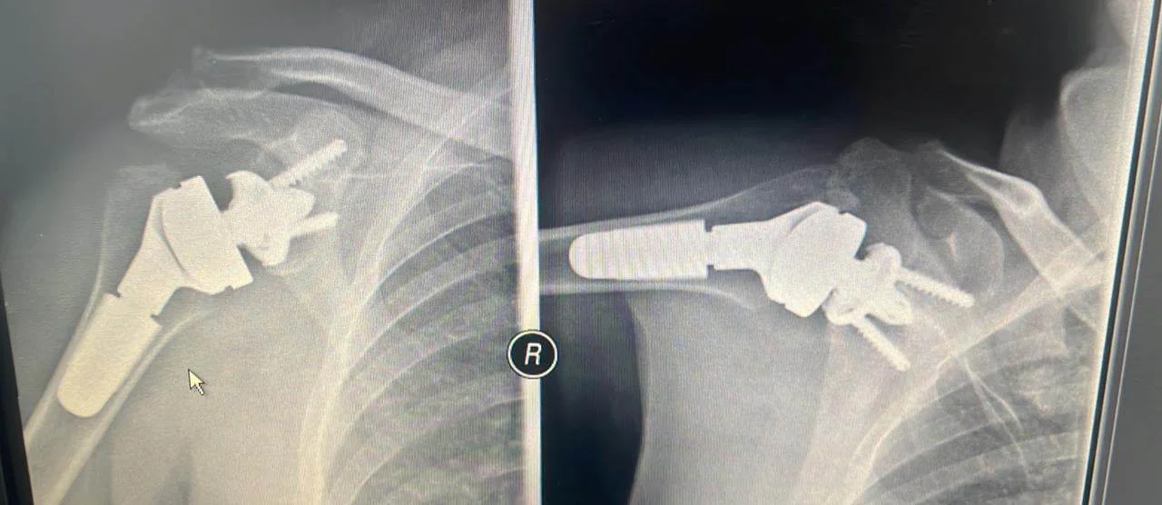
The 59-year-old patient was facing severe pain in her shoulder and arm, accompanied by critical stiffness in the affected area. As she characteristically stated, “I couldn’t use my arm at all, and it hurt all day and every single night.”
One of the first actions taken was compiling a thorough medical history. This was followed by a meticulous clinical examination of the patient, as well as imaging tests (specialized X-rays, CT, and MRI scans). Ultimately, it was determined that the patient suffered from severe deforming arthritis with bone deformities.
The need to completely relieve the patient from pain led to three alternative solutions:
- Conservative treatment, consisting of successive injections and painkillers.
- Anatomical (conventional) total shoulder arthroplasty.
- Reverse total shoulder arthroplasty.
The first option was rejected because such a heavy analgesic regimen could not permanently relieve the patient from her chronic pain. The treatment outcome would only be temporary, mainly due to the advanced stage of the arthritis. Between the second and third options, the therapeutic solution of reverse total arthroplasty was ultimately chosen due to the presence of deforming arthritis along with significant bone deformities and defects.
Pre-operative Planning
The pre-operative planning was highly methodical and precise. The patient’s 3D CT scan, analyzed via specialized computer software, aided in accurately determining the exact joint replacement positioning and the implantation of the various prostheses. It also played a crucial role in selecting the proper prosthesis size, as well as the optimal angle and specific trajectory through which the components would be inserted. This is an extremely critical step, as it ensures maximum precision, speed, and efficacy during surgery, and represents a plan that is always entirely personalized for each individual patient.
The Procedure
The patient therefore underwent a reverse total shoulder arthroplasty, which involves the implantation of artificial surfaces into the shoulder joint positioned in a reversed configuration.
More specifically, the metallic hemispherical ball that replaces the anatomical head of the humerus bone is affixed to the glenoid (the socket of the scapula). This component is initially stabilized with a powerful central screw as well as specialized peripheral screws. Next, a stem is inserted into the humerus bone. These elements lock together using a polyethylene plastic liner. In this manner, we achieve an exceptionally robust and stable new shoulder, which permits movement in every direction and is capable of bearing loads. Biomechanically, the joint’s center of rotation shifts downward and inward, thereby improving the tension of the deltoid muscle, as well as the adjacent tissues, soft structures, and small tendons of the shoulder (collectively known as the rotator cuff).
Surgical Outcome
Indeed, the surgery was completed with absolute success. The patient was discharged the following day and was completely pain-free within a few hours.
It is also worth noting that the patient experienced zero blood loss, while her recovery was rapid and entirely devoid of post-operative pain. She was capable of managing basic self-care within the first 24 hours and utilized a shoulder sling for approximately 10 days, during which she could already move her arm in all directions.
Her immediate return to daily life was facilitated by the small incision, as the method is minimally invasive. This means that during the surgical approach, muscles and tendons are not cut, and the joint replacement is performed without traumatizing the surrounding tissues—a factor of immense importance for patients suffering from severe arthritis and intense daily pain.
Several weeks post-surgery, the patient is ecstatic, feeling that she has fully returned to her normal routine, possessing a complete range of motion, strength, and functionality with absolutely no pain.
Advantages of the Method
The fundamental advantages of this specific method are its minimally invasive nature—requiring only a small incision—and that it is painless and bloodless. A complete elimination of pain is observed within the very first hours following surgery.
Reverse total arthroplasty is performed under general anesthesia, though it can also be carried out under regional anesthesia if the patient prefers, lasting approximately 45 to 60 minutes. Complications range from minimal to non-existent. The surgeon’s operative experience plays a decisive role in this. Consequently, even patients who present as highly challenging cases return to normalcy very rapidly.
The latest, most advanced generations of reverse shoulder arthroplasties are continuously gaining ground and outperforming conventional total arthroplasties in cases of severe deforming arthritis, fractures, acute and chronic dislocations with bone loss and comminution, revisions of previous failed procedures, as well as massive rotator cuff tears and secondary cuff tear arthropathy. Today, reverse total arthroplasty stands as the primary method of choice. At least 80% of shoulder arthroplasties worldwide are reverse total arthroplasties due to their exceptional survivorship and functionality.
A Few Words About the Surgeon
Dr. Ioannis Polyzois and his team are proud to have recently implanted the first reverse total shoulder arthroplasty in Greece using the Arthrex type with MGS Glenoid. This system has demonstrated the best long-term outcomes in Europe and the United States over the last 15 years and features state-of-the-art materials along with a cutting-edge pre-operative planning method that allows for a fully customized implant tailored to the specific patient for the best possible results.
Due to his extensive clinical and professional track record abroad, following a 25-year tenure in Great Britain—including 10 years in a permanent Consultant position—Dr. Polyzois has performed over 9,500 successful surgeries involving the shoulder and upper limb.