Preparation and Modern Preoperative Assessment
The success of the operation begins long before we enter the operating theatre. The preoperative assessment must be methodical, strict, and detailed.
Before the operation, you will need to undergo a series of examinations. Specialised X-rays, magnetic resonance imaging (MRI), and — most importantly — three-dimensional computed tomography (3D CT) are our tools. With the help of a specialised computer simulation programme (3D Preoperative Planning), I map your bone wear with millimetric precision.
This technology allows me to test virtually various sizes of prostheses, to select the ideal angle of inclination and the angle of insertion of the materials. This step is critical: it ensures maximum stability of the implants, protects the existing bone, and drastically reduces the duration of the operation, minimising the strain on your body.
In parallel, in cooperation with our team’s Anaesthesiologist, the most appropriate anaesthesia protocol is chosen, while your medical history, the medication you are taking, and full blood and cardiology examinations are thoroughly reviewed.
How is the surgical operation performed?
Reverse arthroplasty is performed under general anaesthesia, often combined with regional anaesthesia (interscalene block) for the complete elimination of postoperative pain. Its duration is usually around 60 to 90 minutes. In revision cases, where the removal of old materials is first required, the time may be longer.
Using Minimally Invasive Surgery techniques, the approach is made through a small, aesthetically acceptable incision, without unnecessary cutting or injury to the adjacent muscles and tissues.
The steps include:
- Removal of the damaged, arthritic head of the humerus.
- Preparation of the glenoid and placement of the metal spherical implant (the new “ball”), which is stabilised with a powerful central screw and additional peripheral screws for complete integration with the bone.
- Placement of a metal stem inside the humerus, on which a special plastic insert (polyethylene) is fitted, forming the new “socket”.
- Reduction of the joint, which now locks perfectly in its new reverse configuration, ensuring stability and excellent movement.
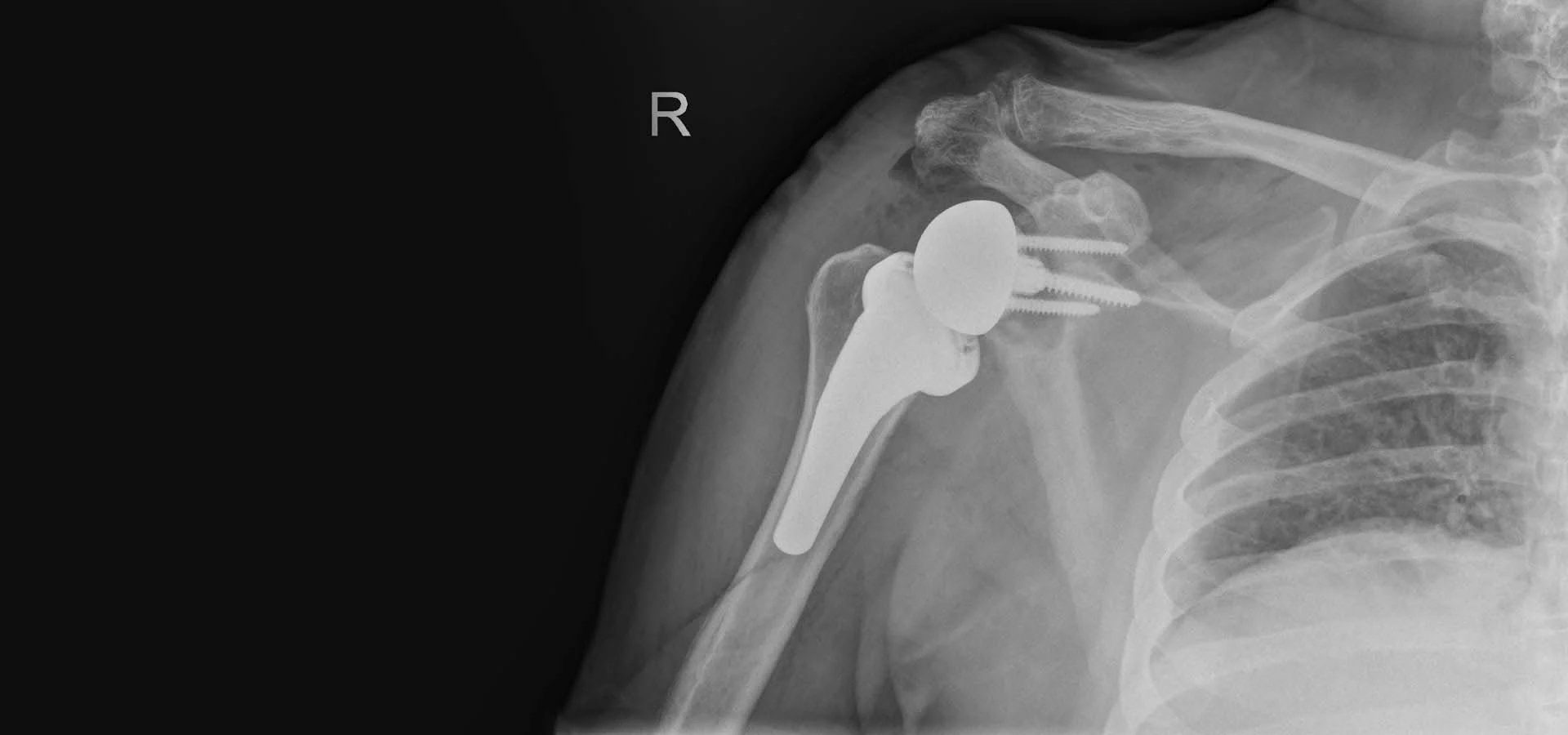
Case Study: Personalised Reverse Total Right Shoulder Arthroplasty
To help you better understand the impact of the operation, let us examine a real case from my practice.
A 59-year-old woman visited me complaining of unbearable pain in her right shoulder, which radiated throughout the arm. The stiffness was so severe that, as she characteristically told me: “I cannot use my arm at all, I cannot comb my hair or get dressed, and I am in relentless pain all day and every night”.
On clinical examination, an inability to actively raise the arm was identified (pseudoparalysis). A thorough imaging assessment with X-rays, CT, and MRI revealed the extent of the problem: the patient was suffering from severe deforming arthritis, accompanied by chronic, massive tear of the rotator cuff and significant bone deficits in the scapula.
We had three options:
- Conservative treatment (injections, painkillers): Rejected, as due to the advanced destruction of the joint, this solution would have been merely a temporary masking of the symptoms.
- Conventional total arthroplasty: Rejected, because the absence of the rotator cuff tendons would have made the conventional prosthesis unstable and non-functional.
- Reverse total arthroplasty: The only definitive, medically indicated option.
Based on the three-dimensional plan (3D CT Planning), we precisely studied her bone deformities. The operation was designed so that the prosthesis would be placed in the ideal position to make maximum use of her healthy bone.
The operation and the outcome:
The surgical operation was completed with absolute success. The procedure was practically bloodless. What was impressive is that the patient was fully relieved from the chronic, agonising pain within just a few hours after recovery from anaesthesia. She was discharged the very next day.
She was able to take care of herself (for basic needs) from the first 24 hours. She wore a light arm sling for protection for about 10 days. Because a minimally invasive technique was used (without cutting muscles), her recovery was impressively quick. Today, weeks after the operation, she has fully returned to her daily life, enjoying an excellent range of motion, renewed strength, and above all, a life without any trace of pain.