Εξάρθρημα ώμου

Shoulder Dislocation

ΠΕΡΙΕΧΟΜΕΝΑ

The shoulder joint is a marvel of human biomechanics, offering us an unprecedented range of motion that no other joint in the body possesses. It allows us to lift the arm overhead, rotate it, throw, hug, and play sports with remarkable speed and strength. However, in medicine and in nature, nothing comes without a price. This extreme flexibility “sacrifices” inherent stability, making the shoulder the most prone joint in the human body to dislocations.

As an Orthopaedic Surgeon, my approach to shoulder instability is strictly research-based and sceptical. The medical literature and clinical practice teach us that a dislocation is never simply a “pop-out” that “went back in”. It is a complex traumatic event that often leaves behind irreparable damage. I do not rely on superficial diagnoses or on a simple X-ray showing the shoulder back in place. I double-check the data, study the MRIs and CT scans myself, and seek out the true extent of the tissue damage. My aim, through this comprehensive and detailed guide, is to explain to you with absolute scientific clarity why the shoulder dislocates, what damage is caused, why recurrence is so frequent, and how modern minimally invasive surgery can return you to a life without the fear of pain.


Understanding the Anatomy and Instability: How the Shoulder Works

To understand the pathology of dislocation, it is of vital importance to take a look at how the shoulder is constructed. The shoulder girdle consists of three main bones: the humerus (the long bone of the arm), the scapula (at the back of the thorax), and the clavicle (at the front).

The main joint of the shoulder, the glenohumeral joint, is often compared to a golf ball balancing on a small, shallow tee.

  • The “ball” is the head of the humerus.
  • The “socket” is the glenoid of the scapula.

The mechanical problem lies in the fact that the surface of the humeral head is 3 to 4 times larger than the corresponding surface of the glenoid, which is almost flat and very shallow. In reality, only 1/3 of the head articulates and is in contact with the glenoid at any given moment and in any movement.

Due to this bony asymmetry, the joint relies exclusively on the soft tissues for its stability:

  • The Labrum: A strong, fibrous ring that surrounds the edge of the glenoid, deepening the socket like a “suction cup” (suction cup effect) by 50%.
  • The Joint Capsule and Ligaments (Static Stability): A very powerful system (capsule) that contains at least three main glenohumeral ligaments, which function as safety belts.
  • The Rotator Cuff (Dynamic Stability): A group of four tendons and muscles that surround the joint and continuously pull it towards the centre of the socket when we move the arm.

When this perfectly coordinated system collapses under the application of extreme force, the head of the humerus separates from the glenoid. If the head dislocates and goes back in immediately (within fractions of a second), we speak of a subluxation. If the ball remains permanently outside the socket and the arm loses its connection with the skeleton, this is a complete Shoulder Dislocation.

Ο μόνος Ορθοπαιδικός στην Ελλάδα στην χειρουργική ώμου και άνω άκρου με προϋπηρεσία μόνιμης διευθυντικής θέσης (substantive Consultant ) για 10 συναπτά έτη στην Μεγάλη Βρετανία

How and Why does the Shoulder Dislocate? Causes and Mechanisms of Dislocation

Shoulder dislocation is not a random condition. It usually happens under specific conditions and is divided into two large categories: traumatic and atraumatic instability.

Traumatic Shoulder Instability

This is the most common cause (it constitutes 95% of cases). It happens after the application of a sudden, violent force that is greater than the strength of the shoulder ligaments. The mechanisms include:

  • Falling on the outstretched arm: The classic mechanism. The arm is in abduction and external rotation (as if we are throwing a ball), and the force of the fall pushes the head violently forwards (Anterior Dislocation).
  • Athletic Collisions: Direct collisions in contact sports (such as football, rugby, martial arts, wrestling, or basketball).
  • Road Traffic Accidents: Acceleration/deceleration forces in car or motorcycle accidents can violently dislocate the shoulder, often causing concomitant bony injuries.

Epileptic Seizures or Electric Shock: In these cases, the extreme and uncontrolled contraction of the body’s own muscles is so strong that it pulls the head out of the joint, usually causing the rarer Posterior Dislocation.

Atraumatic Instability (Ligamentous Laxity)

There is a category of patients in whom the shoulder dislocates (or subluxates) with little or no force, such as during sleep, when putting on a jacket, or reaching out the arm. This type of instability is found in people who are genetically extremely flexible (have “losse” joints). Generalised ligamentous laxity makes the shoulder capsule very “spacious”. It is often observed in swimmers (due to overuse and stretching of the tissues), gymnasts, dancers, or in people with connective tissue syndromes (such as Marfan or Ehlers-Danlos syndromes).

Risk Factors: Who is at Greater Risk?

Through the thorough study of statistics and the literature, we have identified specific factors that dramatically increase the risk of dislocation:

  • Age (15 to 30 years): This is the highest-risk group. Young people are more active, take part in more dangerous activities, and have tighter, more resistant ligaments which, when they break, break “violently”, leading to detachment of the labrum.
  • Sex: Men are twice (or even three times in some studies) as likely to suffer a shoulder dislocation compared to women, mainly due to their participation in high-impact sports.
  • History of Previous Injury: The greatest predictive factor for a dislocation… is a previous dislocation. Once the ligaments are torn, the shoulder becomes vulnerable.
  • Anatomical Particularities: Dysplasia of the glenoid (a congenitally even shallower socket) facilitates the slipping out of the head.

Symptoms: Acute Dislocation and Chronic Instability

The clinical picture depends on whether the patient is experiencing the first, acute episode or suffers from chronic, recurrent instability.

Symptoms in the Acute Shoulder Dislocation:

  • Extreme Pain: It is one of the most painful traumatic events. The patient is unable to move the arm and keeps it stuck to the body, supporting it with the other hand.
  • Tearing Sound: Often the patient reports the sensation or hearing of a loud sound (like a “pop” or “crack”) at the moment the ligaments tear.
  • Visible Deformity: The shoulder loses its normal rounded contour. A characteristic depression (the “epaulette sign”) appears under the acromion, while the head is palpable in front, close to the armpit.
  • Swelling and Spasm: Intense swelling and uncontrolled muscle spasm, as the muscles try in vain to pull the joint back into place.
  • Neurological Symptoms: Numbness, tingling (“pins and needles”) in the arm or fingers, and weakness, suggesting pressure or stretching on the nerves of the brachial plexus (most commonly the axillary nerve).

Symptoms in Chronic Shoulder Instability (Recurrent Dislocations):

When the shoulder has dislocated many times, the initial pain often decreases, but the problems multiply.

  • Sense of Apprehension: The patient lives with the constant fear and insecurity that the shoulder will dislocate. They instinctively avoid specific movements (such as reaching for something on the back seat of the car or throwing a ball).
  • Recurrences with the Slightest Trigger: The shoulder may dislocate even during sleep if the patient turns over, or with a simple, non-violent movement.
  • Chronic Pain: Dull, deep pain in the shoulder, indicative of the progressive wear of the cartilage.

The Importance of Concomitant Injuries: Bankart, Hill-Sachs, and Bone Deficits

In medicine, the accuracy of treatment depends on the understanding of the damage. A dislocation almost always causes significant destruction within the shoulder:

  • Bankart Lesion: This is the most classic injury. As the head dislocates forcefully forwards, it “tears off” and rips the anterior-inferior labrum of the glenoid, along with the ligaments that attach there. If this labrum is not reattached (sutured), the “protective wall” of the shoulder has fallen, and the head is free to slip out again.
  • Hill-Sachs Lesion: As the head of the humerus dislocates and “hangs” outside the joint, the back of it impacts forcefully against the hard edge of the glenoid. This causes a dent, a “compression fracture” (like a crater) on the humeral bone. If the dent is large, every time the arm rotates outwards, this crater “engages” the glenoid, causing a new dislocation (engaging Hill-Sachs).
  • Bony Glenoid Deficits (Bony Bankart): Each new dislocation grinds away and removes precious bone from the glenoid. If the deficit exceeds 20–25% of its total surface, the shoulder is doomed to chronic instability, and heavier, open operations involving bone grafts are required (such as the Latarjet procedure).
  • Other Injuries: In patients over 40 years of age, the dislocation does not usually tear the labrum, but the tendons of the rotator cuff, or causes a fracture of the greater tuberosity of the humerus. This radically changes the treatment plan. Furthermore, the risk of the early development of severe shoulder arthritis increases dramatically.

Diagnosis and Evaluation: The Sceptical, Scientific Approach

As I continuously emphasise, the diagnosis “the shoulder had dislocated” is not enough. We must know exactly what this event left behind. After we reduce the shoulder (put it back in place with special manoeuvres in the emergency department, usually under sedation so that the patient does not suffer), our research work begins at the practice.

I take a thorough medical history (how it dislocated, how many times, whether it went back in spontaneously or at the hospital). Then, I perform a specialised clinical examination. I check your general ligamentous laxity (Beighton criteria) and perform special tests (such as the Apprehension test, the Relocation test, and the Load and Shift test) to feel with my own hands the degree of instability.

Imaging is the final, indisputable arbiter. I personally read and analyse your examinations:

  • Specialised X-rays: In addition to the simple views (Face), I request specialised projections (such as Y-scapular, Axillary view, or Stryker Notch) to see the exact position of the head and any fractures.

Magnetic Resonance Imaging (MRI) or Magnetic Arthrography: This is the “gold standard” for soft tissues. It allows us to see the tear in the labrum (Bankart), the capsule, the ligaments, and the tendons with incredible detail. If the MRI is performed within a few days of the dislocation, the blood in the joint functions as a natural contrast medium. In chronic conditions, I request an MRI with intra-articular contrast for maximum accuracy.

  • Three-dimensional Computed Tomography (3D CT Scan): The ultimate examination if I suspect bony damage. In chronic instabilities, the 3D CT gives us the exact calculation of the glenoid bone deficit as a percentage. From this figure depends whether arthroscopy will succeed or whether we need a different technique.

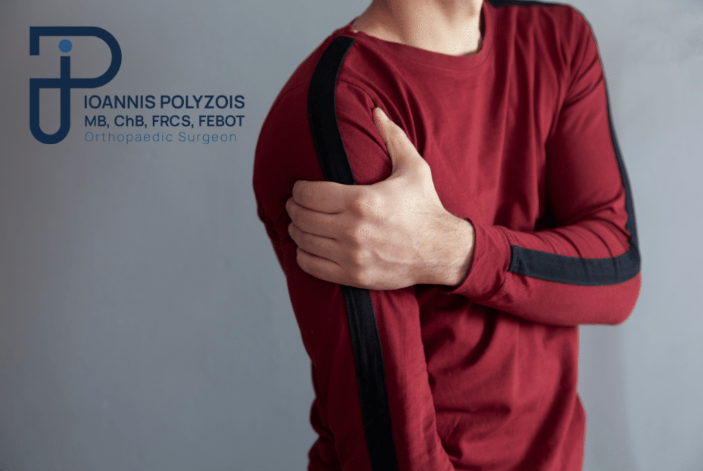
Management: Conservative Treatment vs. Surgical Repair

The dilemma between conservative and surgical management can only be resolved based on scientific data. The international orthopaedic literature (and numerous multicentre studies) demonstrates an inexorable truth:

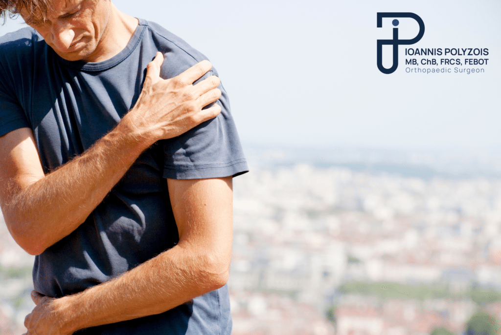
The lower the age at which the first dislocation occurs, the geometrically greater the probability that the patient will suffer a second dislocation. For a young, active patient under 20–22 years of age who plays sports, the probability of dislocating the shoulder again, regardless of how good their physiotherapy is, approaches 80–100%. Each new dislocation further destroys the tissues, “eats away at” the bone, and increases the risk of future, painful arthritis.

Conservative Treatment

We usually recommend it for older patients (over 40–45 years of age), with a low level of athletic/manual activity, provided there are no tendon tears, or for patients with atraumatic, generalised laxity where the problem is the constitution of the tissues.

The protocol includes:

  • Immobilisation in a special sling with the arm often in a neutral or slight external rotation for 2–4 weeks, to allow an initial healing of the torn tissues.
  • Cryotherapy and Non-Steroidal Anti-Inflammatory Drugs (NSAIDs) for pain control.
  • Long-term, aggressive Physiotherapy programme. The goal here is dynamic stabilisation: since the ligaments have stretched out, we train the rotator cuff and scapular muscles to “grip” and keep the joint in place.

The Definitive Surgical Solution: Arthroscopic Shoulder Stabilisation

Provided that the data (age, sports activity, injuries) argue in favour of surgery, my advice is clear: Do not delay. Do not allow your joint to be destroyed with every new dislocation.

The surgical method I apply offers a permanent, reliable, and definitive solution, following the strict GIRFT philosophy (Get It Right First Time). The method of choice is Shoulder Arthroscopy (Arthroscopic Bankart Repair / Stabilisation).

Having a vast volume of cases (high-volume surgeon) and the experience of thousands of operations, arthroscopy in the hands of our team is minimally invasive, bloodless, and extremely safe.

  • The Procedure: Through 3 microscopic openings (4 millimetres), I insert the High Definition camera and the state-of-the-art fine instruments into the shoulder. I identify the exact damage to the labrum.
  • The Repair: Using special, latest-generation bioabsorbable anchors (often knotless anchors) that are placed inside the glenoid, I fix (suture) the torn labrum and the ligaments back into their anatomical position. In parallel, I plicate (tighten) the capsule to “tighten” the shoulder. If there is a Hill-Sachs lesion, I may apply the Remplissage technique (filling the bone defect with a tendon).
  • The Time and the Anaesthesia: The operation lasts about 30–45 minutes. Our specialised anaesthesiologist applies local (regional) anaesthesia (interscalene block) which guarantees that you will not feel any pain for the next 12–24 hours. Light sedation or a short general anaesthesia is applied so that you are completely comfortable.
  • The Recovery: No hospitalisation is required. The patient is discharged and returns home on the very same day. The materials used are 100% biocompatible, integrate with the bone, and are not rejected.

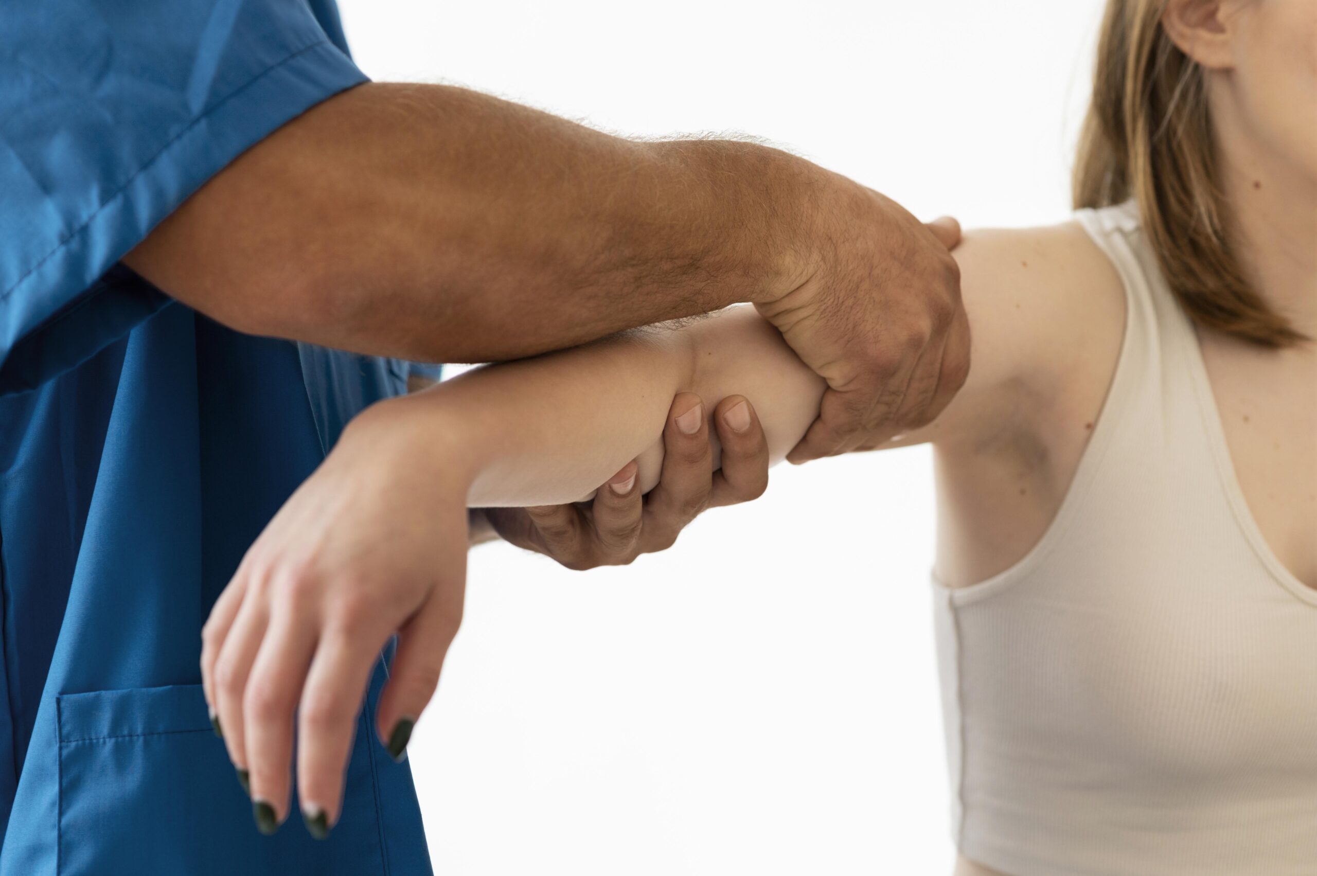
Rehabilitation and Physiotherapy: The Return to Movement

The surgical operation is only half the journey. The other 50% of success — the recovery of full range of motion, strength, and confidence in the shoulder — belongs to strict and structured rehabilitation. I am categorical about this: the seamless cooperation of doctor, patient, and physiotherapist is the cornerstone.

Through our extensive network of specialised physiotherapists throughout Greece, we apply a personalised protocol, which includes:

  • Weeks 1–4 (Protection): The shoulder is protected in a special sling. External rotation is forbidden in order to protect the repair (the anchors “bond” with the bone). Free movement of the wrist and elbow and mild passive exercises (pendulum) are allowed. You can take care of yourself from Day 1.
  • Weeks 4–8 (Mobilisation): The sling is removed permanently. Passive and active-assisted range of motion begins. The goal is to recover elasticity without putting pressure on the sutures.
  • Weeks 8–12 (Strengthening): Isometric and progressive strengthening of the rotator cuff muscles (use of resistance bands) and stabilisation of the scapula begins. You now have full, comfortable movement overhead.
  • Months 4–6 (Return to Sport): The final stage. It includes plyometric exercises, proprioception, and special training depending on the sport. At 4 to 6 months, full and unrestricted participation is allowed even in the most demanding contact sports (e.g. rugby, wrestling) with complete safety.

Meet the Doctor: Dr. Ioannis Polyzois

The treatment of dislocation and the delicate art of arthroscopic stabilisation do not allow for amateurism. They require deep knowledge of anatomy and absolute specialisation. As an Orthopaedic Surgeon with exclusive, targeted specialisation in the conditions, complex trauma, and sports medicine of the shoulder and upper limb, my primary goal is to offer you a definitive, safe, and scientifically documented solution.

Having served as a permanent Consultant in Orthopaedics in the National Health Service of Great Britain (NHS) for more than 10 years, I have managed, at the largest trauma centres in London, the most complex and difficult cases of shoulder instability. My extensive years of further training (fellowships) at the top centres of arthroscopic surgery worldwide enable me to apply the most advanced international techniques in Greece.

To date, I have performed more than 9,000 arthroscopic and open surgical operations. This vast, documented surgical experience — which is reflected in the trust and the hundreds of excellent reviews from our patients — makes me the most competent doctor for the successful execution of such demanding operations, from a simple Bankart repair to complex bone reconstructions.

Every patient is unique, which is why our medical approach is always entirely personalised. We are here to listen to your problem, to resolve every question with honesty, and to design together your return to action.

Cost and Price: Arthroscopic Shoulder Stabilisation

One of the first and most reasonable questions of patients, especially of young people and their families, concerns the financial aspect of the surgical treatment. It is important to understand that shoulder arthroscopy is a medical procedure of high technological specialisation. As such, it is not offered in the form of standardised “packages”.

The final price is shaped strictly on the basis of the needs of your particular joint. The main factors that determine the final cost are:

  • The Extent of the Damage: A simple stabilisation with two bioabsorbable anchors has a different cost from an extensive tear that requires four anchors, or a concomitant tendon repair (e.g. Remplissage).
  • The Cutting-Edge Materials: At our practice, we do not compromise on quality. We use exclusively certified, top materials and ultra-high-strength anchors (usually from America or Switzerland), which guarantee the longevity of the result.
  • The Hospital of Choice: The expenses of the day clinic (one-day surgery), the use of the High-Definition equipment, and the medication.
  • Your Insurance Coverage: The possibility of using the public insurance fund (EOPYY) or your private insurance policy (which often covers 100% of the operation).

Our commitment is to absolute transparency. The exact, honest, and final cost, without any hidden charges whatsoever, is calculated and discussed in detail with you exclusively after the scheduling of an appointment, the clinical assessment, and the study of your MRI/CT examinations by the doctor.

Movement is life!

The fear of instability and the pain of dislocation should not deprive you of the ability to participate actively in life, sport, and the activities you love. Modern orthopaedics has the knowledge and the minimally invasive technology to safeguard your joint.

Contact the doctor today to schedule a thorough diagnostic appointment. We will investigate your data with scientific accuracy, we will resolve every question of yours with honesty, and we will design the most specialised and permanent orthopaedic solution, fully tailored to your needs.

Η κIνηση εIναι ζωH!

Επικοινωνήστε με τον γιατρό για εξειδικευμένη ορθοπαιδική φροντίδα, προσαρμοσμένη στις ανάγκες σας

Συχνές ερωτήσεις

Can a shoulder dislocation occur without intense trauma?

Yes. Although 95% of cases are traumatic (falls, sports), people with generalised ligamentous laxity (hyperelastic joints) may dislocate their shoulder, making a simple movement, such as putting on a jacket, stretching in their sleep, or reaching their arm backwards, without any particular force being applied.

What is the difference between a shoulder dislocation and a subluxation?

In a dislocation, the head of the humerus comes completely out of the glenoid socket and remains there. Medical intervention (reduction) is required for it to return. In a subluxation, the head partially comes out of the socket (slips at the edges) but returns to its normal position on its own, usually within fractions of a second, causing a sudden sensation of instability and pain (“dead arm”).

Can a shoulder dislocation affect daily life in the long term?

If it is not properly treated, the answer is affirmative. Chronic instability (a shoulder that “gives way” or dislocates easily) negatively affects quality of life. The patient lives in constant fear, abandons their favourite sports, has difficulty sleeping, and faces an increased risk of developing early osteoarthritis due to the continuous friction and wear of the bones.

When should immediate medical help be sought after a shoulder injury?

If, after a fall or injury, you feel extreme pain, see a visible deformity in the shoulder, are unable to move the arm, or feel numbness in the fingers, you must immediately go to the nearest hospital (Emergency Department). The shoulder must be reduced quickly by a doctor to reduce the pressure on the vessels, nerves, and cartilage. Never try to “put the shoulder back” yourself or with the help of friends, as this can cause comminuted fractures or neurological injury.

If I dislocate my shoulder once, am I doomed to dislocate it again?

Your age plays the most important role. If you are 18–20 years old and play sports, the risk of repeat dislocation (recurrence) approaches 90–100% despite conservative treatment, due to the way the labrum tears in young ages. By contrast, if the first dislocation occurs at the age of 50, the risk of recurrence is extremely low (below 15–20%), and attention usually turns to the healing of the tendons of the rotator cuff.

How long will I wear the sling after the arthroscopy?

The usual protocol, which ensures the safe integration of the sutures and the labrum with the bone, requires the use of the sling for 3 to 4 weeks. However, during this period, you will remove the sling in a controlled manner several times a day, under our instructions, in order to mobilise the elbow and to perform simple everyday movements (self-care, eating, using a keyboard).

When can I return to the gym and to weights?

The return to exercise is progressive. Running (cardio) and cycling are allowed relatively early (6–8 weeks). The start of exercises with light resistance and bands for the shoulder usually begins in the 2nd–3rd month, always under the guidance of the physiotherapist. The return to full weightlifting in the gym without restrictions is recommended after the completion of 4 to 6 months from the operation.

What is the Latarjet procedure and when do you choose it instead of arthroscopy?

When the patient has dislocated the shoulder very many times (chronic instability), continuous friction “erases” (absorbs) the front bone of the glenoid. If this bony deficit is huge (over 20–25%), simple arthroscopic suture of the soft tissues (Bankart) will fail and the shoulder will dislocate again. In these strictly selected cases, we proceed to the Latarjet procedure, an open surgical technique, where we transfer a piece of bone from the coracoid process (together with its tendons) and screw it in front of the glenoid, creating a powerful, new bony “barrier” with excellent success rates.