Ασβεστοποιός Τενοντίτιδα

Calcific Tendinitis of the Shoulder

ΠΕΡΙΕΧΟΜΕΝΑ

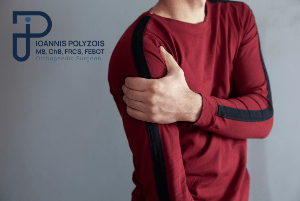
The shoulder joint is an extremely complex and sensitive biomechanical structure, responsible for the greatest range of motion in the human body. However, this complexity makes it vulnerable to conditions that can literally paralyse our daily life. One of the most painful, sudden, and often misunderstood conditions of the region is Calcific Tendinitis of the Shoulder.

As an Orthopaedic Surgeon, my medical philosophy is not based on quick conclusions. I approach every symptom and every condition as a researcher. I am sceptical by nature towards obvious answers, and I double-check every imaging examination and every clinical finding. I know well that in medicine, I am not always infallible at first glance — and neither should you, as patients, rely on the first internet search. Both of us, doctor and patient, must seek the absolute accuracy and the truth that lies behind the pain.

In this exhaustive and fully documented guide, we will place Calcific Tendinitis “under the microscope”. We will analyse the mechanism by which it develops, the causes, its dramatic symptoms, and the most modern, definitive solutions that medicine offers, from conservative management to advanced arthroscopy.


What exactly is Calcific Tendinitis?

To understand the disease, we must look inside the shoulder. The shoulder moves and is stabilised thanks to a group of four important tendons, the well-known rotator cuff. The most important and most often overstressed of them is the tendon of the supraspinatus muscle, which passes through a narrow tunnel (the subacromial space) before attaching to the bone.

Calcific Tendinitis is a condition in which crystals of calcium salts (mainly calcium phosphate) form and accumulate within the very mass and fibres of these tendons. This condition has absolutely no relationship with your diet, your intake of calcium from dairy products, or osteoporosis. It is a localised, biological, and chemical process of “metaplasia”, where the cells of the tendon begin, erroneously, to behave like cells producing bone. This accumulation creates a foreign body inside the tendon, which, depending on the phase it is in, resembles in texture either hard chalk or thick toothpaste. This foreign body causes extreme inflammation, swelling of the tendon, and mechanical friction (catching) against the bones during movement.


How and Why Does the Calcium Form? (The 3-Stage Cycle)

Calcific tendinitis is not a static condition. It follows a very specific biological cycle (known in medicine as Uhthoff’s cycle), which explains why the pain appears so suddenly. This cycle is divided into three main stages:

Pre-calcific Stage

In this early stage, due to micro-injuries or reduced blood supply (hypoxia) in the area of the tendon, the normal cells of the tendon (tenocytes) begin to transform into cells resembling cartilage (fibrocartilaginous metaplasia). In this phase, there is no real calcium, and the patient is usually completely asymptomatic. You feel no pain.

Calcific Stage

This is the stage where calcium begins to be deposited. It is divided into two sub-phases:

Resting Phase: The calcium has formed and resembles hard chalk. The patient may feel a slight discomfort or “catching” in the shoulder, but the pain is tolerable. The condition may remain in this phase for months or even years.

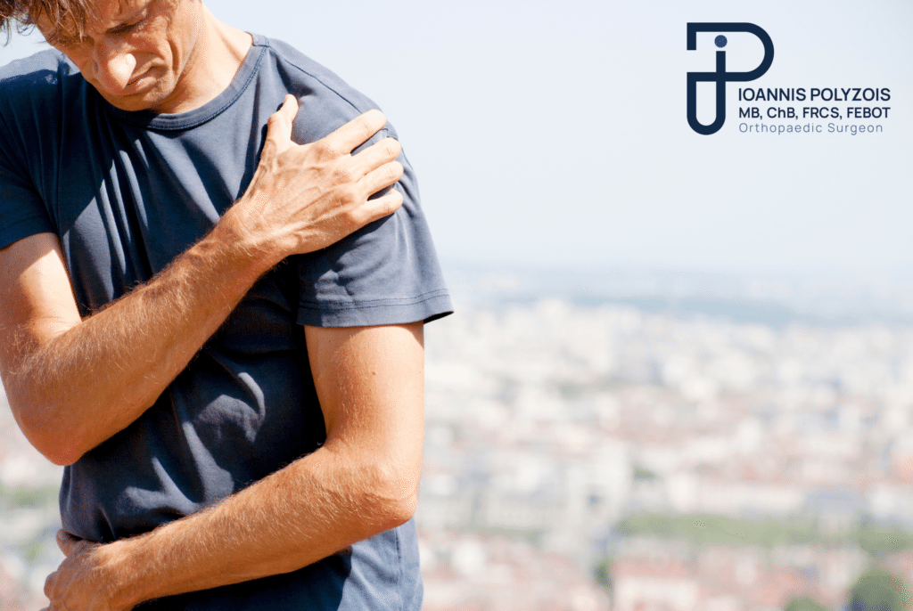
Resorptive Phase: This is the most critical and painful phase. For unknown reasons, the body’s immune system “wakes up”, recognises the calcium as a foreign body, and launches a massive inflammatory attack to dissolve and absorb it. The calcium is transformed into a liquid, viscous mass (like toothpaste) under enormous pressure within the tendon. This chemical inflammation causes one of the most acute, dramatic, and unbearable pains in orthopaedics.


Post-calcific Stage

If the body manages to fully absorb the calcium (something that does not always happen), the tendon enters the phase of healing. New fibroblasts rebuild the tendon. The pain gradually subsides and the shoulder returns to its normal function.

Ο μόνος Ορθοπαιδικός στην Ελλάδα στην χειρουργική ώμου και άνω άκρου με προϋπηρεσία μόνιμης διευθυντικής θέσης (substantive Consultant ) για 10 συναπτά έτη στην Μεγάλη Βρετανία

Causes: What triggers Calcific Tendinitis?

Despite extensive research, the exact primary cause (aetiopathogenesis) of calcific tendinitis remains one of the riddles of medicine. It is not caused by dietary factors or by a simple blow. However, through my scientific research and clinical practice, we have identified very specific predisposing factors:

  • Local Hypoxia (Reduced Oxygenation): The area of the supraspinatus tendon where calcium most often appears has, by nature, a very poor blood supply (the so-called “critical zone”). The lack of oxygen appears to trigger the transformation of the cells.
  • Micro-injuries and Overuse Syndromes: Repetitive micro-traumas from overhead movements cause wear. These injuries cause local swelling of the tendon and increase its pressure and friction inside the subacromial space.

Endocrinological and Metabolic Factors: Studies demonstrate the strong link of the condition with systemic diseases. Patients suffering from Diabetes Mellitus or Thyroid Disorders (hypothyroidism) face a multiplied risk of developing it, as these metabolic disorders affect the microcirculation of blood and the quality of the tendons.

  • Subacromial Impingement Syndrome: The narrowness of the space under the acromion (due to anatomy or osteophytes) rubs the tendon, creating the appropriate inflammatory ground for the deposition of calcium.

Who is more prone to developing the condition?

If we analyse the demographic data, calcific tendinitis “prefers” specific groups of the population.

  • Age: It usually appears in active adults, in the productive age between 30 and 60 years. It is very rarely seen in children or in the very elderly (over 75), where the simple degenerative tear of the tendons predominates.
  • Sex: Research and clinical practice confirm that women are much more prone to developing the condition compared to men (in a ratio of almost 2 to 1), a fact possibly related to hormonal factors still under study.
  • Profession/Activity: Although it can appear in anyone, people who do office work (computer) develop the condition just as often as manual workers or athletes (e.g., tennis, swimming), suggesting that the biological predisposition may be stronger than mere mechanical stress.

The Dramatic Symptoms of the Condition

As previously mentioned, during the majority of the time of its formation, this specific condition can be insidiously asymptomatic. However, when the resorption phase begins, the clinical picture is explosive.

The main symptoms include:

  • Sudden and Extreme Pain: The pain appears suddenly, usually within a period of just a few hours, without any preceding blow. It is piercing, deep, and is often described as “pain as if a knife is stabbing”.
  • Distressing Nocturnal Pain: The inflammation peaks during the evening hours. The result is that the pain wakes the patient up, making sleep in any position practically impossible.
  • Complete Stiffness and “Pseudoparalysis”: The execution of even the simplest shoulder movement is impossible. The patient keeps the arm stuck to the body (as if it is broken) because the slightest lifting increases the pressure on the calcium, causing unbearable pain. The pain may radiate towards the neck, or as far as the elbow and fingers.

The Diagnostic Approach: The Value of Accuracy

A correct diagnosis requires the insight, knowledge, and experience of a specialised Orthopaedic Surgeon. When a patient enters the practice unable to move the arm and reporting sudden, extreme pain without injury, the clinical suspicion of calcific tendinitis is very high.

However, as a sceptic, I never rely on suspicions. The process we follow is strictly protocolised:

  • Medical History and Clinical Examination: I evaluate your medical history in detail. I look for signs of impingement, check reflexes and muscular strength, ensuring that the pain comes from the shoulder and not from a herniation in the neck.
  • The Simple X-ray: In contrast to what many believe, the “gold standard” for the diagnosis of calcific tendinitis is NOT the MRI, but the digital X-ray! The calcium is extremely dense, like bone, and appears dazzlingly white (like a cloud) on the X-ray. I can calculate its size (often 1 to 2 centimetres) and its exact position.
  • Ultrasound: An excellent supplementary tool. It allows us to see the calcium in real time and to examine the flow of blood (with Doppler) around it, evaluating the degree of inflammation.
  • Magnetic Resonance Imaging (MRI): Requested for further evaluation and double-checking. The calcium on the MRI appears as a “black hole” (because it does not contain water). The main reason I request an MRI is to establish the condition of the muscles and tendons of the rotator cuff, ruling out the case of a simultaneous massive tear of the tendon.

Conservative Treatment of Calcific Tendinitis

When the pain is acute, my first and main concern is the rapid relief of the patient. Despite the dramatic picture, 70–80% of cases are initially treated successfully without surgery.

The conservative armoury includes:

  • Rest and Recovery: Avoidance of lifting weights and movements above head height. A sling for 1–2 days may offer relief.
  • Pharmaceutical Therapy: Strong non-steroidal anti-inflammatory drugs (NSAIDs) to control the acute chemical inflammation.
  • Cryotherapy: The application of ice locally significantly reduces the swelling.

Local Injections: The injection of corticosteroid (cortisone) combined with local anaesthetic into the subacromial space (not into the tendon itself, to avoid rupture). This injection is often liberating, “putting out” the fire of inflammation within 24–48 hours.

  • Extracorporeal Shockwave Therapy (ESWT): A modern method where high-energy acoustic waves target the deposit. This causes micro-fragmentation of the calcium and stimulates the blood supply, prompting the body to absorb it more quickly.
  • Needling / Aspiration (Barbotage/Lavage): Under ultrasound guidance, we insert fine needles into the calcium, inject saline solution to dissolve it, and aspirate it, as if cleaning a cyst.
  • Specialised Physiotherapy: Once the acute phase has passed, a targeted exercise programme will restore the range of motion and stabilise the scapula.

Attention: These episodes, in a percentage approaching 15–20%, may be recurrent (relapsing). The patient is trapped in a chronic problem that makes daily life difficult. When this happens, or when the calcium is huge and as hard as stone, conservative treatment reaches its limits.

The Definitive Solution: Shoulder Arthroscopy

When all conservative means have failed, the pain persists for months, and quality of life collapses, modern orthopaedics has in its hands the ultimate “weapon”: Shoulder Arthroscopy.

Arthroscopy is a minimally invasive surgical technique that has revolutionised the treatment of the condition.

How is the operation performed?

Instead of large, painful incisions, the operation is performed through 2–3 microscopic openings in the skin, only 4 millimetres in diameter.

  • Optical Inspection: I insert a special high-resolution camera (the arthroscope) into the shoulder, which projects the interior onto large monitors.
  • Localisation: I examine the supraspinatus tendon. Through a special needle as a guide, I identify the exact location of the calcification. Often, the tendon at that point is swollen like an “angry” volcano.
  • Removal (Debridement): Using microscopic instruments (shavers and forceps), I open the fibres of the tendon. The calcium then bursts out like a “snowstorm” (snowstorm appearance) or like white toothpaste. I clean and aspirate it meticulously, immediately decompressing the tendon.
  • Tendon Repair: If, after the removal of a large volume of calcium, a significant gap (hole) remains within the tendon, I immediately proceed to arthroscopic suturing (repair). I place special, latest-generation bioabsorbable anchors (knotless) and sew up the gap, so that the tendon heals perfectly. Finally, a cleaning (acromioplasty) is performed to create more space for the tendon.

The Advantages of the Arthroscopic Method:

  • Rapid and definitive relief of the patient from the acute pain.
  • No hospitalisation (the patient is discharged a few hours after the operation).
  • Full restoration of the movement and functionality of the shoulder.
  • Minimal trauma to healthy tissues (no muscles are cut).
  • Excellent aesthetic result, without scars.
  • Immediate return to daily life.

Meet the Doctor: Dr. Ioannis Polyzois

The diagnosis of calcific tendinitis and its arthroscopic treatment — especially the correct assessment of whether the tendon needs repair after debridement — require the knowledge and experience of a specialised Orthopaedic Surgeon. An incomplete operation may lead to a future rupture of the tendon.

As an Orthopaedic Surgeon with absolute specialisation in conditions of the shoulder and upper limb, my primary goal is to offer you a safe, definitive, and scientifically documented solution. Having served as a permanent Consultant in Orthopaedics in Great Britain (NHS) for more than 10 years, I have been called upon to manage and treat the most demanding cases.

My extensive further training at the largest centres of arthroscopic surgery worldwide enables me to apply the most modern techniques in Greece. To date, I have performed more than 9,000 arthroscopic and open surgical operations. This vast surgical experience — confirmed by hundreds of excellent reviews from patients who have undergone similar treatments — makes me the most competent surgeon for the successful execution of high-difficulty operations. Every patient is unique, which is why my approach is always entirely personalised. Contact us and forget forever what is troubling you!

Cost and Price: Arthroscopy for Calcific Tendinitis

One of the most natural questions of every patient when surgery is being decided is the financial aspect. Shoulder arthroscopy is a medical procedure of high specialisation and, as such, the cost cannot be answered with a “ready-made”, general price over the phone.

The final price is shaped strictly on the basis of the data of your particular shoulder. Factors that determine the cost are:

  • The Extent of the Operation: A simple arthroscopic debridement and removal of the calcium has a lower cost. If, however, the tendon needs to be “sewn up” (repaired) after debridement, then the cost is added for the bioabsorbable titanium anchors and the special ultra-high-strength sutures.
  • The Materials: We use exclusively top, certified materials from abroad.
  • The Hospital: The expenses of the medical centre (day surgery / one-day clinic).
  • Your Insurance: The use of a public insurance fund (EOPYY) or the coverage by private health insurance.

Our commitment is to absolute transparency. The exact and final cost is calculated and explained to you in full, without any hidden charges, exclusively after the scheduling of an appointment, the clinical assessment, and the study of your imaging examinations at the practice.

Movement is life!

There is no reason to suffer helplessly from the dramatic pain of calcific tendinitis. Modern orthopaedics has the knowledge and the technology to return you quickly to your normal daily life, bloodlessly and definitively.

Contact the doctor immediately to schedule your own diagnostic appointment. We will investigate the causes of your pain with scepticism and accuracy, designing the most specialised orthopaedic care, fully tailored to your needs.

Η κIνηση εIναι ζωH!

Επικοινωνήστε με τον γιατρό για εξειδικευμένη ορθοπαιδική φροντίδα, προσαρμοσμένη στις ανάγκες σας

Συχνές ερωτήσεις

What is calcific tendinitis?

It is an extremely painful condition of the shoulder caused by the accumulation of calcium crystals (salts) within the tendons of the rotator cuff, usually in the supraspinatus muscle. This calcium acts like a “thorn” or “stone” inside the tendon, causing intense inflammation, swelling, and friction during the movement of the joint.

How does calcific tendinitis form?

The exact mechanism remains partially unknown, but we know that it is not related to diet. It is caused by a biological change (fibrocartilaginous metaplasia) of the cells of the tendon, often triggered by poor blood supply (local hypoxia), micro-injuries, overuse, as well as systemic metabolic factors such as the thyroid or diabetes mellitus.

What are the most basic symptoms?

The most characteristic symptoms are acute, sudden, and extremely piercing pain in the shoulder. This pain is aggravated by the slightest movement (difficulty performing movements), while it peaks at night (nocturnal pain), making sleep impossible. It often causes “pseudoparalysis” of the arm, where the patient is completely unable to lift it.

Is it related to the calcium I consume in my diet?

Absolutely none. It is perhaps the most frequent myth. The calcium in dairy products or dietary supplements protects your bones. It does not cause, nor does it worsen, calcific tendinitis. You do not need to change your diet to treat it.

How is calcific tendinitis treated?

Treatment always begins conservatively, especially in the acute phase of pain. It includes rest, strong anti-inflammatory drugs, local cortisone injection (which often brings immediate relief), shockwave therapy (Shockwave/ESWT), and physiotherapy. If the calcium is large, is not absorbed, and the pain persists with relapses, the definitive solution is minimally invasive shoulder arthroscopy for its immediate surgical debridement.

How much does shoulder arthroscopy cost?

The cost is adjusted according to the severity of the condition, the consumable materials (e.g. anchors if a repair of the tendon is required after debridement), the clinic chosen, and your insurance coverage. The detailed cost estimate is always given following a medical appointment and an evaluation of your file.

Is there a chance that the calcium will re-form after the operation?

In the overwhelming majority of cases (over 95%), when the calcium is removed arthroscopically and the subacromial space is properly cleaned, the condition is definitively cured and never returns to the same shoulder.

Is physiotherapy needed after the arthroscopy?

Yes, postoperative rehabilitation is an integral part of success. Although the acute pain of the calcium disappears immediately, the joint and the tendons need a special, progressive programme of kinesiotherapy and strengthening to regain their full strength and elasticity.