How common is elbow bursitis?
Elbow bursitis is particularly common in orthopedic clinics. Statistics show that approximately 65% of cases are non-septic (non-infectious) bursitis and are mainly due to injuries or chronic strain. The remaining 35% represents cases of infection (septic), which require much more aggressive management.
What are the symptoms of elbow bursitis?
The clinical picture of bursitis is usually clear, but the intensity of the symptoms depends directly on its type (aseptic vs. septic). The basic symptoms of elbow bursitis include:
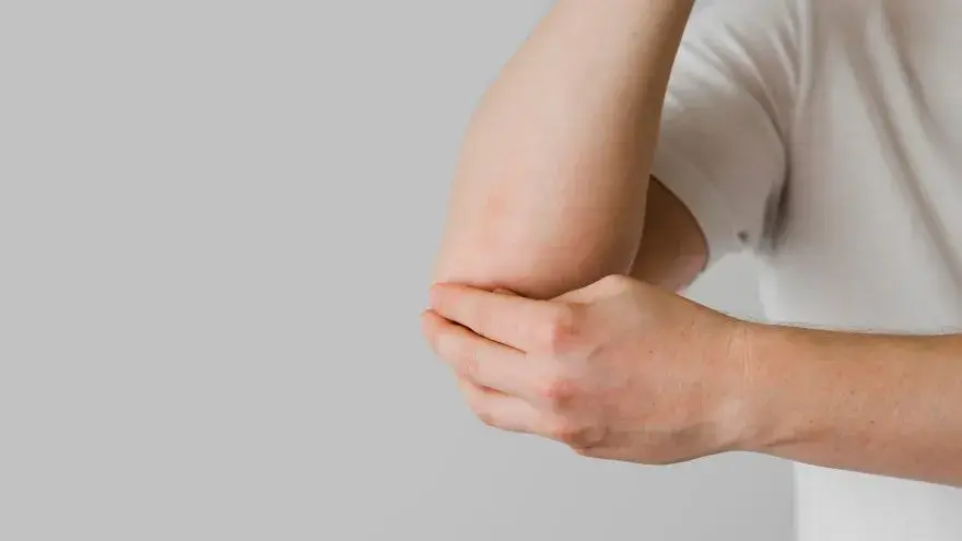
- Swelling and edema around the elbow: It is the first and most obvious symptom. Sometimes, the swelling develops gradually over the passage of weeks and the patient might not notice it until it takes large dimensions (like a ball at the back). Because the skin at the back of the elbow is very elastic, the bursa can fill with a large amount of fluid before it even causes pain.
- Pain and stiffness: As the bursa grows and distends, it begins to press on surrounding nerves and tissues. The pain can be mild in aseptic forms, but becomes sharp during flexion (when the skin stretches). This leads to reduced movement (stiffness) of the elbow, with the patient being unable to fully extend or bend their arm.
- Increased temperature and redness: If the skin around the swelling is red, warm to the touch, and exceptionally tender (even to a light touch), this is the classic warning sign that the bursitis has been caused by an infection (septic). In severe cases, the infection can cause systemic symptoms as well, such as chills, malaise, enlarged lymph nodes in the axilla, and fever. In this case, you must seek medical help immediately.
- Night pain: Especially when the patient is unable to lie down or sleep on the side where the affected elbow is located, due to the local pressure exerted on the mattress.
How is the diagnosis performed?
Accurate diagnosis is the key to deciding the correct therapeutic approach. The specialized orthopedic surgeon, Mr. Polyzois, will see you at his clinic, take your specific history (e.g., what occupation you do, if there was a recent injury or scratch on the skin), and examine you carefully. The clinical picture on its own often “betrays” the condition, but the differential diagnosis between aseptic and septic is of vital importance.
If Mr. Polyzois suspects that the bursitis is septic or has been caused by an infection, or if he wants to rule out other underlying causes for your elbow pain, further examinations might be needed, such as:
- Plain X-ray of the elbow (X-rays): Although the X-ray does not show soft tissues or the fluid of the bursa, it is necessary to rule out conditions such as calcific tendinitis, bone spurs (osteophytes) that might be injuring the bursa internally, or a fracture of the olecranon, as well as osteoarthritis.
- Ultrasound and Magnetic Resonance Imaging (MRI): These imaging examinations show soft tissues with absolute clarity. They are used to confirm the size of the bursa and, primarily, to rule out the extension of an infection to adjacent tissues, soft tissues, tendons, or the bone (osteomyelitis).
- Blood check: Blood tests (such as CRP and ESR) can reveal markers of inflammation in the body, while checking uric acid helps in the diagnosis of gout.
- Fluid aspiration (Puncture) from the bursa: It constitutes the most important diagnostic examination. The doctor uses a thin needle to remove (aspirate) some fluid from the swollen bursa. The fluid is sent to a specialized laboratory for microbiological culture and analysis. If the fluid is yellowish and clear, it is usually an aseptic inflammation. If it is cloudy, purulent, or contains crystals, it confirms infection or gout respectively.
How is olecranon bursitis treated?
The way in which elbow bursitis is treated depends strictly on whether it is caused by an infection or not.
Conservative Treatment (For Aseptic Bursitis)
If from the examination and puncture it emerges that no infection exists (aseptic), treatment is initially conservative and aims at reducing inflammation and absorbing the fluid:
- Rest and Activity Modification: The most important step. Avoidance of the activity causing the bursitis and strict prohibition of resting the elbow on hard surfaces. Frequently, the use of special protective “cushions” or elbow pads adjusted for the elbow is recommended.
- Ice Therapy: Application of ice (never directly on the skin, but wrapped in a towel) for 15-20 minutes, several times a day. The cold causes vasoconstriction and drastically reduces edema and pain.
- Medications (NSAIDs): Non-Steroidal Anti-Inflammatory pills or ointments help significantly in reducing the pain and internal inflammation of the bursa.
- Temporary Immobilization: In more persistent situations, stopping the movement of your elbow with a simple splint or elastic bandage for a short period of time can prevent the further production of fluid.
- Aspiration and Cortisone Injection: If the bursitis persists and does not present improvement with the above within three to six weeks, Mr. Polyzois can fully aspirate the fluid from within the bursa (relieving the pressure immediately) and, at the same time, make a local injection of cortisone inside the now empty sac. Cortisone is an exceptionally powerful anti-inflammatory that suppresses the irritation of the bursa’s cells, preventing the reappearance of the fluid. After the injection, a tight bandage is applied.
Treatment of Infection / Septic Bursitis
Septic bursitis does not subside on its own and, if left without treatment, bacteria can infiltrate the blood (septicemia) or the bones.
In case of infection, Mr. Polyzois will administer antibiotics (usually by mouth in mild cases, or intravenously in the hospital in more severe ones). It is of vital importance for the patient to take the antibiotics for the entire recommended period, even if symptoms disappear. Parallelly, the doctor will aspirate your bursa with a syringe (perhaps more than once over a period of days) to remove as much of the purulent fluid as possible, which will be sent for analysis to the laboratory, so it is confirmed that the absolutely appropriate antibiotic is administered. If infectious bursitis does not respond quickly to antibiotics, urgent surgical drainage is required.
Surgical Treatment (Bursectomy)
Surgical intervention becomes necessary in three main cases:
- When a septic bursitis is not controlled with medications.
- When the bursa has undergone chronic wear, has thickened excessively, and is full of fibrous tissue (chronic incurable aseptic bursitis).
- When the fluid returns continuously (recurrences) despite repeated punctures and cortisone injections.
Mr. Polyzois, as a specialized Director Elbow Surgeon, offers the patient a definitive solution to the problem with a 100% success rate if conservative treatment fails.
The operation (called a bursectomy) is entirely bloodless and painless. The specialized anesthesiologist administers regional anesthesia (fully numbing only the arm) and thus the patient can remain awake throughout the entire duration (Awake Surgery), if they desire so, of course, avoiding general anesthesia.
During the operation, the entire pathological and inflamed bursa is removed. At the same time, any other lesions are corrected, such as an accompanying tendinitis or local osteoarthritis (e.g., bone spurs on the olecranon bone that might have been injuring the bursa internally are “filed down”) and an extensive debridement is done. The body, over the passage of months, will create on its own a completely new, healthy bursa in the place of the old one, which will function smoothly without inflammations.
Postoperative course: Rehabilitation is rapid. The patient is discharged from the hospital (Day Clinic) a few hours after the completion of the operation. A light bandage is placed and the patient moves their elbow immediately without any substantial restriction, starting physiotherapy. The result, both functionally (complete absence of pain and unhindered movement) and aesthetically (elimination of the ugly swelling), is excellent.