Κατάγματα Ώμου

Shoulder Fractures

ΠΕΡΙΕΧΟΜΕΝΑ

The shoulder joint, thanks to its unparalleled freedom of movement, allows us to perform everything from the most delicate and demanding tasks to the most dynamic sporting activities. However, when an accident leads to injury and fracture of the bones that comprise it, the pain, the weakness, and the disruption of daily life are dramatic. Shoulder fractures represent one of the most frequent and, at the same time, most complex orthopaedic challenges.

As a doctor and scientist, my approach to a fracture is far removed from standardised, “ready-made” solutions. Medicine is a continuous search for the truth. I remain sceptical towards easy diagnoses, I research in depth, and I double-check every piece of information, every X-ray, and every symptom. At my practice, the treatment decision is a collaborative process: we study the data together, we discuss the options, and although we know that in medicine there are no dogmas, we always try — with the greatest possible accuracy — to choose the path that will return you to a normal life.

In this exhaustive and fully detailed guide, we will examine every aspect of shoulder fractures, from the complex anatomy and the mechanisms of injury, to the most modern surgical techniques of osteosynthesis and arthroplasty.

The Complex Anatomy of the Shoulder Girdle

To understand the seriousness and the treatment of a fracture, we must first “map out” the area. The shoulder girdle is not a simple bone, but a complex system. It consists of three main bones, which must function in absolute harmony:

The Humerus (Head and Proximal Portion)

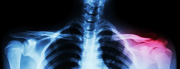
It is the long bone of the arm. Its upper part (the proximal humerus) ends in a spherical structure, the head, which is the “ball” of the joint. Just below the head, there are two bony protrusions, the greater and lesser tuberosities, to which the powerful tendons of the rotator cuff attach. A fracture here — which is also the most common — does not simply break the bone, but destabilises the entire muscular system of the shoulder.

The Scapula

This is a large, triangular, and relatively flat bone located at the back of the thorax. The scapula constitutes the “base station” of the shoulder and, in turn, consists of distinct, critical parts:

  • The Glenoid: The concave portion (the “socket”) with which the head of the humerus articulates, forming the main glenohumeral joint.
  • The Acromion: A bony protrusion that constitutes the “roof” of the shoulder.
  • The Coracoid Process: A small, beak-shaped bone at the front, critical for the attachment of important ligaments and muscles (such as the biceps).
  • The Body of the Scapula: The main flat portion.

All of the above areas can be involved in comminuted or hairline fractures, making each case unique.


The Clavicle

The elongated bone that connects the sternum to the acromion of the scapula. It functions as a “strut” or beam, keeping the shoulder away from the thorax. Clavicle fractures are extremely common, especially in young people and athletes.

Ο μόνος Ορθοπαιδικός στην Ελλάδα στην χειρουργική ώμου και άνω άκρου με προϋπηρεσία μόνιμης διευθυντικής θέσης (substantive Consultant ) για 10 συναπτά έτη στην Μεγάλη Βρετανία

How are Shoulder Fractures Caused?

The causes differ radically depending on the age group and the physiology of the patient. They are mainly divided into two large categories:

Low-Energy Injuries (Osteoporotic Fractures)

These are most often seen in older patients (usually over 65 years of age). Due to osteoporosis, the quality and density of the bone are reduced. A simple fall from body height — for example, tripping at home or on the pavement — and the instinctive support on the outstretched arm or the direct impact of the shoulder on the ground is enough to shatter the head of the humerus.

High-Energy Injuries

These mainly concern younger, active individuals. Their bone quality is excellent, so to cause a fracture, an enormous force is required. Such situations include:

  • Road traffic accidents (especially with a motorcycle).
  • Falls from great height (e.g., work accidents, fall from a ladder).
  • Serious sporting accidents (cycling, skiing, martial arts, extreme sports).

In these cases, the fractures are often accompanied by dislocation (fracture-dislocation) or by very severe injuries to the underlying soft tissues, i.e., tears in the tendons and ligaments, even injuries to nerves (such as the axillary nerve) or vessels.

Symptoms: How will I understand that I have suffered a fracture?

The clinical picture of a shoulder fracture is usually unmistakable and leaves no room for doubt:

  • Acute, piercing pain: The pain appears immediately at the moment of the fall and is dramatically aggravated by the slightest attempt at movement.
  • Complete inability to move: The patient usually keeps the injured arm stuck to the body with the good arm to protect it (characteristic position).
  • Swelling and deformity: The shoulder loses its normal, rounded shape. Especially in clavicle fractures, you can see the bone protruding under the skin.
  • Extensive ecchymosis (bruising): In the following hours or days, a large bruise usually descends from the shoulder towards the arm, the elbow, or even to the thorax, due to the internal bleeding from the broken bone.
  • Numbness or tingling: If the fracture has pressed on or injured the nerves of the area, the arm may feel numb or “heavy”.

The Importance of an Accurate Diagnosis: The Philosophy of the Double Check

This is precisely where the difference between a hasty and a flawless scientific approach lies. In shoulder fractures, the first action in the emergency department is always X-rays (at least in two or three different planes). However, the anatomy of the scapula and the head is three-dimensional, and a two-dimensional X-ray can easily “hide” a serious displacement or comminution.

I am not satisfied with a simple image. As a doctor who researches, I insist on the performance of a Three-Dimensional Computed Tomography (3D CT Scan) for every fracture that presents the slightest suspicion of displacement or comminution. The CT scan gives us a three-dimensional “map” of the bone, showing exactly how many pieces it has broken, where the tuberosities have been displaced, and how stable the area is.

In certain cases, if I suspect a simultaneous tear in the tendons (especially in younger patients or in cases of dislocation), I may also request Magnetic Resonance Imaging (MRI). The diagnosis is not simply the identification of the fracture, but the full understanding of its “personality”. Only with the correct data can we make the correct therapeutic decision.

Conservative Treatment: When do we avoid surgery?

The good news is that a very large percentage of shoulder fractures (about 70–80% of proximal humerus fractures) are hairline or minimally displaced. The broken pieces remain anatomically close to one another, held in place by the periosteum, the capsule, and the tendons.

In these cases, the management is conservative (non-surgical) and includes:

  • Immobilisation: Placement of the arm in a special sling, usually for 3 to 6 weeks, depending on the progress of healing.
  • Analgesic Therapy and Cryotherapy: Targeted pharmaceutical therapy for the control of pain during the first difficult weeks.
  • Strict Monitoring: Repeated X-ray check every 1–2 weeks to make sure that the fracture has not “slipped” or shifted secondarily.

Personalised Physiotherapy: Conservative treatment does not mean indifference. One of the biggest mistakes is the absolute immobility of the shoulder for months, which leads to a “frozen shoulder”. We start immediately, under my guidance, with special passive movements (such as pendulum exercises) to keep the joint alive, gradually adding active exercises as the bone “sets” (consolidates).

Surgical Treatment: The Modern Options

When the fracture is significantly displaced, comminuted (broken into 3, 4, or more pieces), open (the bone has pierced the skin), or when there is a coexisting vessel injury, surgical operation is the only way. If it is not reduced (placed back in position) surgically, the patient will end up with a stiff, deformed limb, full of chronic pain and early osteoarthritis.

Treatment is not “one size fits all”. It is entirely personalised. The surgical options that we have available, thanks to the evolution of biomedical technology, include:

Osteosynthesis (Plates, Screws, and Intramedullary Nails)

The goal here is to “save” and reconstruct your own bone. The technique mainly concerns younger patients with good bone quality.

  • Titanium Plates and Screws (Locking Plates): We use special anatomical plates where the screws “lock” onto the plate, creating an extremely stable scaffold. We reduce (assemble) the broken pieces like a puzzle and stabilise them with the material.
  • Modern Third-Generation Intramedullary Nails: A less invasive method for specific types of fractures. A titanium rod (nail) is inserted into the canal of the bone, offering tremendous mechanical stability with minimal incisions. It is often chosen for fractures of the diaphysis (the middle portion) of the humerus or for specific fractures of the head.

Shoulder Arthroplasty: The Solution for Severe Fractures

But what happens with older patients with severe osteoporosis? In cases where the bone is so shattered that it resembles an “eggshell”, the screws cannot hold. Furthermore, in severe fractures, the blood supply to the head of the humerus is destroyed, leading to necrosis (avascular necrosis). Here, the solution is the replacement of the joint.

  • Shoulder Hemiarthroplasty: We replace only the broken head of the humerus with a metal, artificial spherical implant, preserving your own glenoid. The tendons of the rotator cuff, which are often detached along with the bony fragments, are meticulously sutured around the prosthesis.

Reverse Total Shoulder Arthroplasty: It constitutes the pinnacle of modern orthopaedics for fractures. In the patient with a chronic fracture/dislocation, in those with pre-existing arthritis, or when the tendons are irreparably damaged (rotator cuff tears), conventional replacement will fail because there are no tendons to move the arm.

In the reverse arthroplasty, we reverse the anatomy: we place the concave plastic part on the humerus and the metal ball on the scapula. This shifts the centre of gravity and allows the external muscle (deltoid) to take over all the work of movement, bypassing the need for tendons. In recent years, Reverse Arthroplasty has offered a clearly superior, guaranteed, and immediate functional result compared to conventional methods, eliminating pain and returning the patient to their independence.

Recovery and Physiotherapy: The Day After

Whether we follow a conservative or surgical path, the return to normality requires hard work and excellent guidance. Physiotherapy begins almost immediately.

  • Phase 1 (Protection): During the first weeks (0–6), we protect the osteosynthesis or the prosthesis. The arm rests in the sling, but special passive exercises are performed under the supervision of the physiotherapist.
  • Phase 2 (Mobilisation): As the bone consolidates (6–12 weeks), we begin active-assisted movements, progressively increasing the range of motion of the shoulder. The patient takes care of themselves for basic needs.
  • Phase 3 (Strengthening): After the 3rd month, provided that the fracture has fully healed radiographically, we add strengthening exercises with resistance. The full recovery of a shoulder fracture can last from 6 months to a full year.

Meet the Doctor: Dr. Ioannis Polyzois

In the management of such complex trauma, the experience of the surgeon determines the final outcome. There is no room for half-measures or superficial diagnoses.

As an Orthopaedic Surgeon with absolute specialisation in the conditions, fractures, and arthroplasties of the shoulder and upper limb, my primary goal is to offer you a safe, definitive, and scientifically documented solution to your problem.

Having served as a permanent Consultant in Orthopaedics in Great Britain (NHS) for more than 10 years, I have been called upon to manage, at the largest trauma centres in London, the most demanding, comminuted, and complex cases of fractures. My fifteen years of further training and the performance of many hundreds of operations of osteosynthesis and shoulder arthroplasty enable me to apply the most modern international techniques in Greece.

This vast clinical and surgical experience makes me the most competent for the successful execution of any injury around the shoulder, however simple or complex it may be. Every patient is unique, which is why my approach is always entirely personalised. I am here to listen to your problem, to study your examinations meticulously, and to design together, with accuracy and safety as our guide, your return to a daily life full of movement.

Η κIνηση εIναι ζωH!

Επικοινωνήστε με τον γιατρό για εξειδικευμένη ορθοπαιδική φροντίδα, προσαρμοσμένη στις ανάγκες σας

Cost and Price: Surgical Treatment of Shoulder Fractures

The cost of treating a shoulder fracture is an important and entirely reasonable question. However, just as every fracture is different (from a simple crack that requires a sling, to a comminuted fracture that requires reverse arthroplasty), so too the cost is adjusted according to the medical procedure.

The final amount is determined by parameters such as:

  • The surgical method (Osteosynthesis with plates and screws vs. Intramedullary nail vs. Arthroplasty).
  • The cost of the implants, as we strictly use only materials of top quality and durability, is made of titanium.
  • The hospital or clinic, the time spent in the operating theatre, and the days of hospitalisation (usually 1–2 days for major operations).
  • The coverage through your insurance fund (EOPYY) or your private insurance policy.

The accurate and transparent estimate of the cost, without any hidden charges, is given exclusively after the scheduling of an appointment, the clinical examination, and the detailed evaluation of your imaging examinations. Our goal is your treatment, always with reliability and respect for the patient.

Movement is life!

A shoulder fracture is a serious ordeal, but it should not be the end of your active life. Modern orthopaedics has the knowledge, the technology, and the tools to repair even the most complex injuries.

Contact our practice immediately for specialised orthopaedic care, tailored to your needs. We are here to diagnose the problem with absolute accuracy and to design together your fastest and safest return to health.

Συχνές ερωτήσεις

Can a shoulder fracture permanently affect the strength of the arm?

If the management (conservative or surgical) is the appropriate one and the physiotherapy is done correctly, most patients recover most (if not 100%) of their strength and functionality. In severe comminuted fractures, a small stiffness in the end ranges of motion may remain, but it rarely impedes daily life.

Can I sleep on my side after a shoulder fracture?

For the first 1.5 to 2 months (6–8 weeks), sleeping on the broken or operated shoulder is strictly forbidden, as it may displace the fracture. Sleeping in a semi-sitting position (with many pillows behind the back) or on the back with a supportive pillow under the elbow is the recommended solution.

Is there a risk of developing a “frozen shoulder” or stiff shoulder?

Yes, and it is one of the most frequent complications in fractures. If the shoulder remains completely immobile in the sling for weeks without the correct, controlled passive movements that we prescribe, the capsule contracts and “freezes” (post-traumatic stiffness syndrome). For this reason, proper and timely physiotherapy is equally important as the operation.

Can a shoulder fracture cause pain in the neck or back?

Absolutely. When the shoulder is injured and immobilised, the muscles of the neck (such as the trapezius) and of the back take on all the weight of the arm (compensatory contraction). This often creates muscle spasms and pains around the scapula and the base of the skull, which is managed with light massage, warm compresses on the neck, and physiotherapy.

If I have plates and screws fitted (osteosynthesis), will they set off the airport machines?

Modern osteosynthesis materials and arthroplasties are mainly made of medical titanium or special alloys, which in most cases do not activate metal detectors. If they do, simply showing the surgical incisions or a small medical certificate, which we provide for you, resolves the matter immediately.

Do the osteosynthesis materials (the screws) need to be removed in the future?

As a general rule, if the materials do not bother you, they remain in the body for life. An exception is younger patients or cases where, after the complete consolidation (healing) of the fracture (after 1 to 1.5 years), the plate creates friction with the tendons or the acromion. In these cases, their removal can be scheduled with a much lighter operation.

When can I bathe on my own again?

If you are treated conservatively, you can wash yourself carefully with the help of a relative from the first day, removing the sling, provided that the arm stays hanging and relaxed. If you are operated on, thanks to the special waterproof adhesive dressings that we use, you can shower 2–3 days after discharge, keeping the area clean and avoiding rubbing the incisions.

When will I be able to drive again?

Driving requires the use of both arms and the ability to react suddenly. Regardless of the method of treatment, driving is strictly forbidden for at least 6 to 8 weeks, until the bone shows clear signs of healing on the X-ray and you have recovered the necessary muscular strength.

I am elderly. Is it worth having an arthroplasty, or simply waiting for it to heal?

If the fracture is comminuted and is left without an operation (neglected), the head of the humerus will set in the wrong position, the arm will not lift, and the pain will be permanent due to arthritis. The Reverse Arthroplasty, despite its severity, has changed the picture. It is designed precisely for older patients, allowing them to eat, dress, comb their hair, and take care of themselves again without pain. It is worth every effort.