Specialized Orthopedic Care: Mr. Ioannis Polyzois
The management of upper limb fractures requires excellent training, deep knowledge of microanatomy, and high surgical precision. Orthopedic Surgeon Mr. Ioannis Polyzois guarantees the safest and most effective treatment for every complex injury.
An International Path of Excellence
Mr. Polyzois is the only Orthopedic Surgeon in Greece, specialized in shoulder and upper limb surgery, who held a permanent directorial position (substantive Consultant) for 10 consecutive years in the National Health Service (NHS) of Great Britain. Having successfully managed thousands of complex trauma cases and severe fractures abroad, he applies the most modern international medical protocols, offering his patients a high level of specialized care, aimed at full anatomical restoration and immediate mobilization.
Analytical Presentation of Fractures per Anatomical Region
-
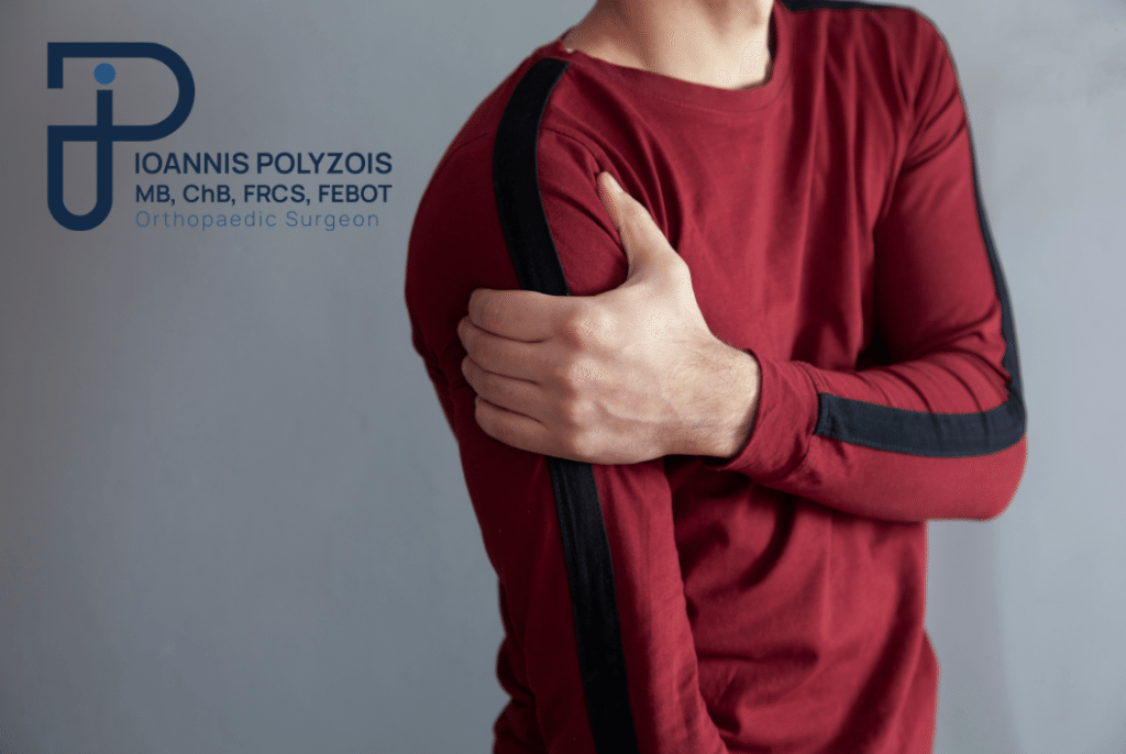
Fractures in the Shoulder Region
-
Clavicle Fractures
They are very common, especially in young people and athletes, and usually occur after a direct fall onto the shoulder (e.g., from a bicycle or motorcycle) or during contact sports. They manifest with severe pain, swelling, bruising, and often a visible deformity (“step”) in the collarbone area.
While most non-displaced fractures are treated conservatively with a special sling (figure-of-eight or simple sling), significantly displaced fractures, shortened fractures, or those with skin tenting require surgical stabilization (internal fixation with anatomical titanium plates and screws) to avoid malunion and permanent functional weakness of the shoulder.
-
Scapula Fractures
These are rare injuries that require high-energy trauma, such as car accidents or falls from a great height. Because the scapula is surrounded by thick muscle groups, these fractures often heal conservatively. However, if the fracture extends into the joint surface (glenoid cavity) or involves the neck of the scapula with significant displacement, surgical repair is necessary to prevent early post-traumatic arthritis and chronic shoulder instability.
Proximal Humerus Fractures (Humerus Head)
Particularly common in elderly patients, especially women with osteoporosis, after a simple fall from standing height. However, they can also occur in young patients due to high-energy injuries. They are classified into fractures of 2, 3, or 4 parts, depending on the number of displaced bone fragments.
Non-displaced fractures are managed conservatively with immobilization and early physical therapy. Displaced and unstable fractures in active patients are treated surgically with osteosynthesis using special locked anatomical plates. In elderly patients with severely comminuted fractures or when the blood supply to the humeral head is completely disrupted (high risk of avascular necrosis), the modern solution is reverse total shoulder arthroplasty, which ensures immediate pain relief and excellent functional restoration.
-
Humerus Shaft Fractures
These involve the middle part of the arm bone and usually result from direct blows or twisting forces. A critical element in these fractures is the close anatomical relationship of the humerus with the radial nerve, which wraps around the bone. A fracture in this area can compress or lacerate the nerve, causing temporary or permanent paralysis (inability to extend the wrist and fingers, known as “drop hand”).
Many shaft fractures can be successfully treated conservatively with special functional braces (Sarmiento brace). Absolute indications for surgical treatment (osteosynthesis with plates and screws or intramedullary nailing) include open fractures, injuries to both arms, non-compliance with the brace, or associated radial nerve palsy that occurs after closed manipulation.
-
Elbow Fractures and Dislocations
-
Distal Humerus Fractures
These are highly complex and demanding intra-articular fractures that occur in both children (supracondylar fractures) and adults. Because they involve the joint surface, perfect anatomical reconstruction is required down to the millimeter.
Treatment is almost exclusively surgical, using dual anatomical locked plates to allow immediate post-operative movement and prevent the elbow’s greatest enemy: permanent severe stiffness.
-
Olecranon Fractures
The olecranon is the prominent tip of the ulna at the back of the elbow, where the powerful triceps muscle inserts. A fracture here is almost always displaced because the triceps pulls the bone fragment upward.
Therefore, surgical intervention is required in the vast majority of cases, either with the tension band wiring technique (for simple fractures) or with anatomical low-profile plates (for comminuted fractures), to restore the elbow extension mechanism.
Radial Head and Neck Fractures
They usually occur after a fall onto an outstretched hand with the elbow extended. They cause pain on the outer side of the elbow and a significant limitation in the rotation of the hand (pronation/supination).
Simple, non-displaced fractures are treated with short-term immobilization and immediate movement. Displaced or comminuted fractures require surgical intervention, which may include micro-osteosynthesis with mini screws/plates, or, in cases of severe comminution where reconstruction is impossible, replacement of the radial head with an anatomical metallic prosthesis (arthroplasty) to maintain elbow stability.
Elbow Dislocations and Complex Injuries
An elbow dislocation occurs when the bones of the forearm are completely separated from the humerus, usually after a violent fall. It is a medical emergency that requires immediate reduction under sedation or anesthesia.
When a dislocation is combined with fractures of the radial head and the coronoid process, it is called the “terrible triad of the elbow”. This is an extremely unstable injury that requires complex surgical reconstruction of both the bones and the torn collateral ligaments to prevent chronic instability and stiffness.
-
Forearm Fractures (Radius and Ulna)
-
Isolated Fractures of the Radius or Ulna Shaft
An isolated fracture of the ulna shaft often occurs from a direct blow when a person raises their arm to protect their head from an attack (known as a “nightstick fracture”). If it is non-displaced, it can be treated with a cast; otherwise, it requires surgical plate fixation.
-
Both-Bone Forearm Fractures
When both the radius and the ulna are broken, the fracture is inherently unstable. Because the parallel arrangement and the relationship between these two bones are what allow us to rotate our palm up and down, any deviation leads to a loss of this movement.
In adults, these fractures are treated exclusively surgically with rigid internal fixation using plates and screws on both bones through separate incisions.
Monteggia Fracture-Dislocation
This is a complex injury consisting of a fracture of the proximal third of the ulna accompanied by a simultaneous dislocation of the radial head at the elbow. It requires accurate surgical fixation of the ulna, which usually leads to spontaneous reduction and stabilization of the radial head.
Galeazzi Fracture-Dislocation
The exact opposite of Monteggia: it involves a fracture of the distal third of the radius accompanied by a dislocation of the distal radioulnar joint at the wrist. It requires rigid surgical fixation of the radius with a plate and evaluation of the stability of the joint at the wrist, which may need temporary stabilization with pins (K-wires).
-
Wrist and Hand Fractures
-
Distal Radius Fractures
This is perhaps the most common fracture in the human body, especially in postmenopausal women due to osteoporosis, but also in young people after high-energy trauma (e.g., snowboarding, rollerblading, or falls). Depending on the direction of the displacement, they are called Colles fractures (displacement backward) or Smith fractures (displacement forward).
If the fracture is extra-articular and can be aligned perfectly, it can be treated conservatively with a cast for 5-6 weeks. However, if the fracture enters the joint surface, is comminuted, or cannot hold its alignment, gold-standard treatment involves open reduction and internal fixation with a modern volar anatomical locked titanium plate. This allows the patient to avoid long cast immobilization and start using their hand within days.
-
Scaphoid and Other Carpal Bone Fractures
The scaphoid is the most common carpal bone to fracture, usually after a fall onto an extended wrist. It is a “treacherous” fracture because it often does not appear on initial X-rays and can be mistaken for a simple sprain. Furthermore, the scaphoid has a peculiar blood supply that enters from its distal end; a fracture at its waist or proximal pole can cut off the blood supply, leading to non-union (pseudarthrosis) or avascular necrosis.
If diagnosed early and non-displaced, it is treated with a specialized cast for a long period. In cases of displacement or to avoid a long time in a cast, percutaneous or open fixation with a special compression screw (headless compression screw) is performed.
Metacarpal and Phalanax Fractures (Fingers)
These are very common injuries resulting from direct blows, sports (e.g., basketball, volleyball), or work accidents. A characteristic fracture is the “boxer’s fracture”, which involves the neck of the 5th metacarpal (below the pinky knuckle) after a punch.
Depending on the displacement, the presence of rotational deformity (when the finger overlaps another during bending), and the stability, treatment ranges from simple splinting or buddy taping to surgical stabilization with micro-plates, mini-screws, or percutaneous pins (K-wires).