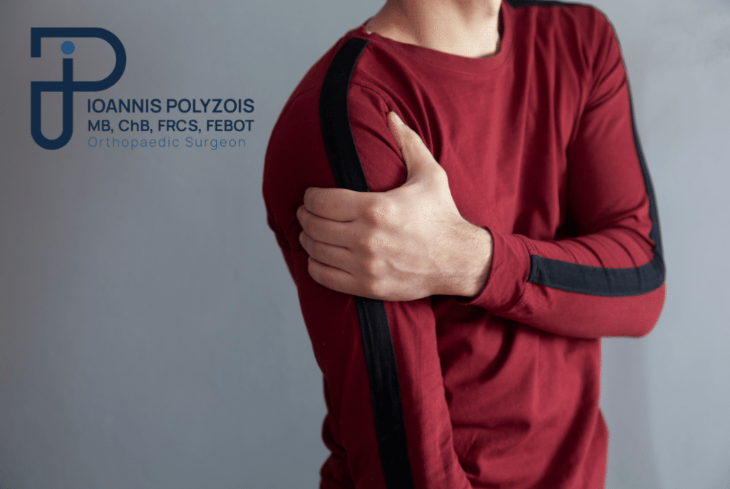
Symptoms of elbow tendinitis
Epicondylitis, unfortunately, is one of the conditions characterized by intense, debilitating, and deep pain, sometimes on the inner and sometimes on the outer side of the elbow, which can worsen with the passage of time. There are cases where the pain is so sharp that it can radiate (travel) along the forearm down to the wrist or hand.
The most common symptoms include:
- Localized Pain: Pain centered on the bone of the elbow, which increases dramatically when you palpate or press the area.
- Sensation of weakness in the wrist and reduced grip: The patient discovers that they no longer have the strength to hold objects. Even holding a coffee mug, a pen, or a pot can become impossible.
- Painful handshake: The simple movement of squeezing someone’s hand or turning a doorknob causes pain that resembles a “sting”.
- Morning Stiffness: A sensation of “catching” and tightness in the elbow in the morning as soon as you wake up, which improves slightly as the joint “warms up”.
- Sensation of numbness in the fingers of the hands: Particularly in medial epicondylitis, due to the proximity of the area to the ulnar nerve, the edema can cause mild pressure translated into numbness.
- Night Pain: In acute phases, the pain can appear even in a state of rest, disrupting sleep.
Diagnosis of elbow tendinitis
Valid diagnosis is the first and most critical step for correct treatment. The diagnosis of elbow tendinitis is made primarily through a thorough clinical examination by the specialized orthopedic doctor.
As in every medical examination, so in this specific one, the patient’s medical history constitutes the “A and Z”. The doctor will ask about your occupation, your sports habits, when the pain started, and which specific movements exacerbate it, in order to recommend the most appropriate treatment method for you.
Clinical Examination
In more detail, the doctor subjects the patient to a local tenderness test, carefully palpating both the inner and outer side of the elbow region.
In combination with the local tenderness test, the doctor will perform a specific diagnostic test on the patient. For example:
- Cozen’s Test (for lateral epicondylitis): The patient makes a fist, turns the wrist upward (extension), and the doctor exerts resistance against them. If this causes sharp pain in the elbow, the test is positive.
- Medial Epicondylitis Test: The patient bends their wrist inward under the doctor’s resistance.
Imaging Examinations
In most cases, the clinical examination suffices. However, in cases where the condition does not respond to treatment or the doctor requires a further picture, it is possible to ask the patient to perform additional examinations in order to exclude the possibility that symptoms originate from some other condition (e.g., arthritis, cervical radiculopathy, nerve entrapment syndrome):
- Plain X-ray (X-ray): It helps to rule out osteoarthritis, any old fractures, or the presence of calcifications (calcium deposits) inside the tendon.
- Ultrasound: An excellent and fast examination showing the thickness of the tendon, the presence of inflammation (hyperemia), and any small tears.
- Magnetic Resonance Imaging (MRI): It is the most detailed method. It is used in persistent, chronic conditions to evaluate with absolute accuracy the degree of tendon degeneration (tendinopathy) or the existence of a larger tear that might need surgical management.
Treatment of elbow tendinitis
Following a thorough examination by the doctor, the patient receives the appropriate personalized treatment in order to stop the progression of the lesion and not suffer henceforth from epicondylitis.
Conservative Management (First Line)
- Rest and Modification: The first step and the most important is for the patient to abstain immediately from the activity that caused the epicondylitis. It does not necessarily mean complete immobility, but avoidance of movements triggering the pain.
- Medication: When the pain is unbearable or quite intense, then the appropriate medication is administered which includes taking simple analgesics and Non-Steroidal Anti-Inflammatory Drugs (NSAIDs), either in the form of pills or in the form of topical gel.
- Ice Therapy: In combination with the medication, ice can be applied (always wrapped in a towel) 3-4 times a day for about 15 minutes each time, especially after activity, to reduce edema.
- Splint – Elbow Strap (Bracing): In cases where avoiding the activity that caused the epicondylitis is deemed impossible (e.g., due to work), the patient can use a specific compression splint for epicondylitis (the elbow strap). This is tied slightly below the elbow, absorbs vibrations, and changes the pull point of the muscles, so that when the patient makes the movement, the inflamed tendon remains protected.
Physiotherapy and Special Exercises
Targeted physiotherapeutic interventions have proven very effective.
- Stretching and Strengthening: Mild stretches (stretching), which are followed by certain eccentric strengthening exercises (where the muscle produces work while lengthening), are the gold standard for the rehabilitation of tendons.
- In order for the implementation of the exercises to be done in the correct way, it is important that they are initially done in the presence of a specialized physiotherapist, so that there are no further injuries to the area.
- Shockwave Therapy: The physiotherapist, apart from monitoring and demonstrating the exercises, can soothe the pain with various modern means, such as for example extracorporeal shockwave therapy. The acoustic waves stimulate blood circulation and activate the natural healing process of the tendons, “breaking” the chronic scar tissue.
Biological Treatments (PRP Injections)
When classic conservative treatment does not yield after a few months, modern orthopedics turns to Regenerative Medicine. PRP (Platelet-Rich Plasma) injections constitute an exceptionally successful solution. A small amount of blood is taken from the patient, centrifuged to isolate the growth factors, and subsequently injected exactly at the spot of the damaged tendon, promoting its rapid biological regeneration without the side effects of traditional cortisone.
Surgical Management
In a small percentage of patients (approximately 5-10%), where pain persists for more than 6 to 12 months and dramatically affects the quality of life despite treatments, surgery is indicated. Modern techniques (such as minimally invasive surgery – MIS or arthroscopy) allow the surgeon to clean the damaged, necrotic tissue of the tendon and improve the blood supply of the area, securing a definitive solution to the problem. The operation is fast (usually Day Clinic) and recovery is rapid.
Prevention of Elbow Tendinitis
Prevention is always better than treatment. To avoid the appearance of the condition, follow these rules:
- Ergonomics at the office: Adjust the height of your chair and desk so that the elbows form a 90-degree angle and the wrists are straight when typing. Use an ergonomic mouse.
- Correct Sports Equipment: If you play tennis or padel, ensure that the thickness of the racket’s grip is correct for your hand and the strings are not excessively tight.
- Warm-up and Stretches: Never start manual work or sports without 5-10 minutes of warm-up and stretches of the forearm muscles.
- Listen to your body: If you feel a “pull” or fatigue in the elbow, stop the activity. Pain is the body’s warning signal.