Shoulder Tendon Tear

ΠΕΡΙΕΧΟΜΕΝΑ

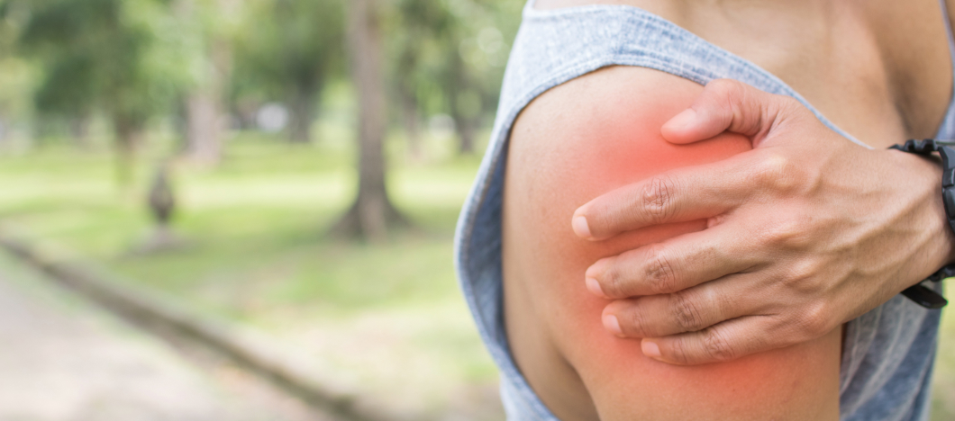
Shoulder pain is perhaps the most frequent problem that I am called upon to treat daily at my practice. If you are here, on drpolyzois.gr, reading these lines, it is very likely that either you or someone close to you is suffering from acute or chronic pain, weakness, and exhausting restriction of arm movement. Perhaps you have already heard the diagnosis that is frightening to many: Shoulder Tendon Tear (scientifically known as a rotator cuff tear).

As an Orthopaedic Surgeon, my philosophy is not based on quick, superficial answers, but on fully evidence-based medicine. I am sceptical by nature towards easy solutions and standardised protocols. It is not enough simply to read the paper of an MRI scan; we must examine the person, understand their daily needs, their biology, and how the pain has altered the quality of their life.

We must be honest: A tendon tear does not automatically and obligatorily mean surgery for everyone. Medicine is the art of correct selection. However, when surgical intervention is required to save the functionality of the limb, modern minimally invasive arthroscopic surgery now offers excellent, permanent, and safe results.

In this exhaustive, analytical guide, we will place the tendon tear under the medical “microscope”. I will explain to you with clarity and scientific accuracy the complex anatomy of the shoulder, the mechanism that leads to the destruction of the tendon, the warning signs, as well as all the modern methods of conservative and surgical treatment to win back the freedom of your movements.

What is a Shoulder Tendon Tear? The Anatomy of Movement

To understand how and why a tendon “breaks”, we must first understand the anatomy that surrounds it. The shoulder is the joint with the greatest, most impressive range of motion in the human body. It functions like a ball (the head of the humerus) balancing on a very shallow socket (the glenoid of the scapula).

Because this bony socket does not, on its own, offer stability, the shoulder relies on a dynamic suspension system: a group of four critical muscles and their tendons, called the Rotator Cuff. These four tendons embrace the head of the humerus like a tight “sleeve” and keep it perfectly centred within the joint, allowing you to lift and rotate the arm with strength.

These four “protagonists” are:

  • Supraspinatus: Located at the top. It is the most important, the most overstressed, and the one that suffers a tear in 90% of cases. It is mainly responsible for the abduction (lifting) of the arm to the side.
  • Infraspinatus: Located at the back and decisively assists in the external rotation of the shoulder (as when you comb your hair).
  • Subscapularis: The largest and strongest, located at the front and responsible for internal rotation (e.g., when you put your hand behind your back).
  • Teres Minor: A smaller muscle at the back that cooperates for external rotation.

When in medicine we say “shoulder tendon tear”, we are referring to the detachment, tearing, or separation of the fibres of the tendon (usually of the supraspinatus) from its anatomical position of attachment on the bone. The “engine”, that is, comes off its mounting, with the result that the transmission of force is lost.

The Types of Rotator Cuff Tears

Tears are not all the same. Depending on the size and the depth of the damage, they are categorised into:

  • Partial-thickness Tear: The tendon has undergone wear, has frayed, or has partially torn (only on the upper or lower fibres), but has not been completely severed. Imagine a thick nautical rope whose external strands have begun to fray, but the rope has not snapped in two. It causes intense pain, but the arm often retains its strength.
  • Full-thickness Tear: The tendon has been severed completely from its upper to its lower surface and has been completely detached from the bone (head of the humerus). In these cases, there is a real “gap” (hole) in the tissue. The muscle contracts and “pulls” the end of the tendon inwards (retraction), creating severe weakness and pain.
  • Massive Tear: The most serious condition, where more than two tendons have been completely severed (e.g., supraspinatus and infraspinatus together). The shoulder loses its functionality, often creating a picture of “pseudoparalysis”.

Ο μόνος Ορθοπαιδικός στην Ελλάδα στην χειρουργική ώμου και άνω άκρου με προϋπηρεσία μόνιμης διευθυντικής θέσης (substantive Consultant ) για 10 συναπτά έτη στην Μεγάλη Βρετανία

The Basic Causes: Why does the shoulder tendon “break”?

A rotator cuff tear is not a condition with one and only one cause. The scientific community categorises it into two large, distinct categories:

Acute Traumatic Tear

It happens suddenly, usually in younger, active people or athletes. In these cases, a completely healthy tendon is subjected to a force that exceeds its breaking limit. It may be caused by:

  • Falling with the entire body weight on an outstretched arm or direct impact on the shoulder.
  • The sudden lifting of a very heavy object (e.g., lifting a piece of furniture) with poor technique.
  • A shoulder dislocation. Particularly in patients over the age of 40–45, a violent shoulder dislocation does not simply tear the ligaments, but very often detaches the tendon of the rotator cuff as well.

Degenerative Tear Due to Chronic Wear

This constitutes the overwhelming majority of cases I treat in my practice. Tendons do not break in one day. With the passage of time and daily use, the tendon wears, just like the belt of a machine that operates nonstop. This is the result of a series of factors:

  • Age-related Degeneration: The biological quality of collagen is reduced. These tears are extremely common in people over 55–60 years of age. Many people in their seventh decade of life have partial tears without even knowing it.
  • Reduced Blood Supply (Local Hypoxia): It has been scientifically demonstrated that a specific area of the supraspinatus tendon (the “critical zone”, about 1–2 centimetres before its attachment to the bone) has, by nature, a very poor blood supply. As we age, the microcirculation is further reduced. Without sufficient blood, the cells of the tendon are unable to heal the normal daily micro-injuries, with the result that the tissue degenerates.
  • Impingement Syndrome: Above the tendons of the shoulder, there is a bony protrusion (the roof), called the acromion. When lifting the arm, the tendon and the bursa pass through a narrow “tunnel”. If this space narrows (due to anatomy or due to the development of osteophytes / “spurs”), the tendon “rubs” and “catches” on the bone. This chronic mechanical friction gradually leads to the thinning and ultimately the severing of the tendon (like a rope that rubs against the edge of a rock).
  • Repetitive Stress (Overuse): Professions (painters, carpenters, electricians, hairdressers) or sports (tennis, swimming, weightlifting) that require the arm to work continuously above head level dramatically increase the risk of a tear.

The Warning Symptoms: How will I understand that I have a tear?

In medicine, nothing is absolute. Not all tears present the same symptoms. Small, degenerative tears that develop over decades may be asymptomatic, as the body finds ways to adapt. However, when the damage extends or becomes acute, the classic warning signs are loud:

  • Pain at rest and especially at night: This is perhaps the most characteristic and distressing symptom of a rotator cuff tear. The patient has unbearable difficulty sleeping on the side of the affected shoulder. The acute, deep pain wakes them repeatedly during the night, causing chronic insomnia and exhaustion.
  • Pain during movement: Particularly when you try to lift the arm to the side or when you perform overhead movements. The pain is also acute when you stretch the arm behind the back (e.g., to fasten a bra, to put the hand in the back pocket, or to reach for something in the back seat of the car).
  • Muscular Weakness: The tearing of the “engine” causes weakness. The arm seems “heavy”. In large full-thickness tears, the patient cannot hold the arm in the air or drop objects.
  • Crepitus and Mechanical Symptoms: A sensation of “clicking”, snapping, or intense creaking when you move the shoulder in specific positions, the result of the friction of the head against the acromion due to the absence of the tendon.

Important, Urgent Note: If you have suffered an injury (e.g., a fall) and you feel sudden, dramatic weakness in lifting your arm to the side, an immediate orthopaedic assessment is required. Acute, traumatic tears have dramatically better outcomes if they are operated on early (within the first weeks), before the muscle retracts, undergoes atrophy, and is transformed into fat (fatty degeneration), a condition that often makes the damage irreversible.

The Diagnostic Approach: Clinical Examination and Imaging

As I continuously emphasise in my practice, we do not treat imaging examinations; we treat people. The diagnosis of a tendon tear requires a research spirit and a comprehensive, strictly scientific approach:

  1. Medical History and Clinical Examination: This is the alpha and the omega. First, I listen to how and when you hurt. Then, through special, targeted clinical tests of kinesiology (such as the Jobe test, Hawkins test, Drop Arm test, Bear Hug test), I isolate and check the strength of each individual muscle separately. A thorough clinical examination often gives me the diagnosis before I even see the MRI.
  2. Simple X-ray: Although the tendon is not visible on X-ray, it is the first step to rule out bone injuries, arthritis, or the formation of large osteophytes (spurs) that cause impingement. If the space between the head and the acromion has narrowed, this is an indirect indication of a large tear.
  3. Shoulder Ultrasound: An excellent, quick, non-invasive, and dynamic examination. Its great advantage is that we can see the tendon “in motion”, pinpointing exactly where it catches.
  4. Magnetic Resonance Imaging (MRI): It constitutes the ultimate diagnostic tool (Gold Standard). I personally read and study the MRI. It reveals to us with crystal-clear accuracy:
    • The exact size of the tear (in centimetres).
    • Whether it is of partial or full thickness.
    • The degree of retraction (how much the tendon has been “pulled” backwards).
    • The quality of the muscle itself. The finding of muscular atrophy or fatty degeneration (when the muscle begins to be replaced by fat due to disuse) is perhaps the most critical information for surgical planning and for predicting the success of the repair.

Conservative Treatment: Can Surgery be Avoided?

The answer is a clear Yes. As a doctor guided by scientific data and respect for the patient, I know and apply the rule that not all tears need surgery.

Age, the needs of the patient, the quality of the tissues, and the type of tear play a decisive role. Older patients (over 70–75 years of age) with chronic, degenerative tears who do not have high physical and sporting demands, or patients with focal partial-thickness tears, very often respond excellently to conservative treatment. The body has amazing ways of compensating (using other muscles to cover the gap).

The conservative, non-surgical plan includes:

  • Modification of Activities: Avoidance of lifting weights, sudden movements, and restriction of activities that require the arms overhead (above the head).
  • Non-Steroidal Anti-Inflammatory Drugs (NSAIDs): For the immediate control of pain and acute inflammation of the bursa (always under medical guidance).
  • Intra-articular Injections:
  • Cortisone: A very powerful anti-inflammatory. It offers dramatic and rapid relief from the nocturnal pain. However, it must be administered with strict medical caution (no more than 2–3 times), as the careless, excessive use of cortisone has been shown to further weaken the fibres of the tendon, increasing the risk of a complete tear.
  • Biological Therapies (PRP – Platelet-Rich Plasma): They are often used in small partial-thickness tears. The purpose is to use the growth factors of your own blood to enhance biological healing.

Specialised Physiotherapy (The most critical element): Even a severed tendon does not mean the end. The physiotherapist will help you strengthen the remaining healthy muscles of the rotator cuff, the deltoid, and the stabilisers of the scapula, so that they take on the “load” of the severed tendon and dynamically stabilise the joint.

Arthroscopic Tendon Repair: The Modern Surgical Solution

When conservative treatment fails after 3–6 months of proper effort, when we are talking about young (under 60), dynamic, and active patients, or when the tear is acute/traumatic and causes significant loss of strength, surgery is the only way. The “leave it and we’ll see” logic with a large tear is dangerous. The persistence of a severed tendon leads, with mathematical accuracy, to the progressive widening of the hole, to the destruction (fatty atrophy) of the muscle, and ultimately, to the development of severe, permanent osteoarthritis of the shoulder (Rotator Cuff Tear Arthropathy).

In modern orthopaedics, the heavy “open” type operation with large incisions and destruction of the deltoid muscle is now in the past. The operation is performed arthroscopically exclusively.

How is the Arthroscopic Repair Performed?

Shoulder arthroscopy is a Minimally Invasive Surgery technique that requires tremendous specialisation.

  • The Approach: Through 3–4 very small openings (about half a centimetre in size) around the shoulder, we insert a high-resolution camera (arthroscope) and special, microscopic surgical instruments.
  • The Preparation (Acromioplasty): The camera projects the interior of the shoulder onto large monitors. Initially, with special radiofrequency instruments and shavers, I clean the inflamed bursa. Then, I “smooth out” and remove the dangerous osteophytes (spurs) from the acromion (Acromioplasty), enlarging the space so that the sutured tendon does not rub and tear again in the future.
  • The Repair (The “Stitching”): The severed tendon is identified, cleaned of dead tissues, mobilised (to overcome the retraction), and brought back to its anatomical position, on the bone of the head of the humerus, which we have prepared.
  • The Anchors: To fix the tendon to the bone, we use special arthroscopic anchors (microscopic screws made of bioabsorbable material or medical titanium). These anchors are screwed into the bone. On their heads, they carry extremely durable, ultra-high-technology sutures, with which we sew the tendon and “tie” it tightly.

Cutting-Edge Techniques: At my practice, I apply the most modern suture techniques, such as the Double-row repair or Suture Bridge, where indicated. With this technique, we cross the sutures, creating a net, ensuring the maximum possible mechanical strength, pressing the tendon evenly onto the bone. This creates the ideal environment for the complete biological healing (“setting”) of the tissues.

The Advantages of Arthroscopy:

  • Safety and Accuracy: Zero injury to the surrounding healthy muscles, as they are not cut to gain access to the joint.
  • Reduced Pain: Minimal postoperative pain thanks to the small incisions and the regional anaesthesia.
  • Immediate Return: Discharge on the same day (Day Clinic). No hospitalisation.
  • Aesthetics: No unsightly scars. The small openings close with a simple adhesive or a single suture.

Postoperative Rehabilitation and Return to Daily Life

I must be absolutely clear with my patients: The operation corrects the mechanical problem, but the biological work must be done by your body. Success depends 50% on excellent surgical technique and 50% on your own disciplined participation in physiotherapy rehabilitation. The tendon needs time, patience, and respect to biologically “set” onto the bone.

Rehabilitation follows a strictly structured, international protocol, which I supervise closely in cooperation with specialised physiotherapists:

  • Phase 1: Protection and Healing (0 – 4 or 6 Weeks) The shoulder is protected in a special brace-sling (with an abduction pillow). Active movement of the shoulder is forbidden, so that the anchors are not pulled out before the tendon sets. Only passive movements are allowed (the physiotherapist moves your arm, without you contracting any muscle) to avoid stiffness. You move the wrist and elbow freely.
  • Phase 2: Active-Assisted Movement (4 – 10 Weeks) The brace is removed permanently. You begin to move your arm by yourself (with the help of the other hand, pulleys, or a stick), gradually recovering the full range of motion of the joint.
  • Phase 3: Muscular Strengthening (10 – 16 Weeks) Now that the tendon has set, the muscle must become strong. The progressive muscular strengthening of the rotator cuff and the scapula begins, initially using isometric exercises, then resistance bands and light weights.
  • Phase 4: Full Return (4 – 6 Months) Completion of rehabilitation and return to heavy manual work and demanding sporting activities (e.g., tennis, swimming) without any fear or restriction.

Meet the Doctor: Dr. Ioannis Polyzois

The arthroscopic repair of a rotator cuff tear is one of the most demanding operations in modern orthopaedics. The knowledge of when, how, and what exactly must be sutured to ensure the viability of the tendon does not allow for amateurism. As an Orthopaedic Surgeon with exclusive, targeted specialisation in the conditions, trauma, and arthroscopic surgery of the shoulder and upper limb, my primary goal is to offer you an absolutely safe, definitive, and scientifically documented solution.

Having served as a permanent Consultant in Orthopaedics in the National Health Service of Great Britain (NHS) for more than 10 years, I was trained in a system that requires excellence and does not accept half-measures. My many years of further training at the top centres of arthroscopic surgery abroad enable me to apply the most advanced international suture techniques (such as the double-row technique) in Greece.

To date, I have performed more than 9,000 arthroscopic and open surgical operations. This vast, documented clinical experience — which is reflected in the trust of hundreds of patients — makes me the most competent doctor for the successful execution of such demanding operations, ensuring an excellent functional result. Every patient is unique to me. At our practice, we do not simply read MRIs; we listen to your problem with honesty and design together your return to normality.

Cost and Price: Arthroscopic Shoulder Tendon Repair

The question about the financial aspect of a surgical operation is entirely reasonable. It is, however, of vital importance to understand that the arthroscopic repair of a tendon is not a “standardised” procedure, and consequently, its cost cannot be given over the phone or via a general price list on the internet. Highly specialised orthopaedic surgery is an entirely personalised medical procedure.

The final cost of the operation is shaped strictly by the specific needs of your particular damage. Factors that determine the price are:

  • The Extent of the Tear: A small tear that requires 1 or 2 anchors to suture has a different cost in consumables from a huge, massive tear that requires 4 or 5 anchors and complex bridging techniques.
  • The Implants (Anchors): At our practice, we make no compromise on your safety. We use exclusively top, certified materials and anchors (bioabsorbable or medical titanium) from the best manufacturers worldwide (USA, Switzerland).
  • The Infrastructure: The expenses of the clinic (One-Day Clinic), the operating theatre, and the use of the state-of-the-art High-Definition arthroscopy systems.
  • Your Insurance Coverage: The use of EOPYY, as well as Private Health Insurance contracts, plays a decisive role, as they very often cover the largest part or all of the medical and hospital expenses.

My basic principle is absolute transparency towards the patient. The exact, honest, and final cost, without any “hidden” charge or surprise, is analysed and discussed in detail exclusively after the scheduling of your appointment, the thorough clinical examination, and the evaluation of your MRI scan at the practice.

Movement is life!

Pain, weakness, and sleepless nights do not need to be a permanent part of your daily life. Modern orthopaedics provides absolutely safe, tested, and definitive solutions, adapted to your biology.

Contact our practice today to schedule a thorough diagnostic appointment. With a strict scientific approach, we will study your examinations, resolve every question with absolute honesty, and together we will organise your recovery plan to return you with strength and health to the life you love.

Η κIνηση εIναι ζωH!

Επικοινωνήστε με τον γιατρό για εξειδικευμένη ορθοπαιδική φροντίδα, προσαρμοσμένη στις ανάγκες σας

Συχνές ερωτήσεις

Can the tendon “set” (heal) on its own without surgery?

We must be absolutely honest: The answer is No. A full-thickness tear of the tendon cannot suture itself, set, or regenerate on its own, because the muscle pulls the severed end of the tendon away from the bone (like a snapped rubber band). Conservative treatment does not “set” the tendon; it simply reduces the pain and trains the remaining muscles to take on the work of the severed one.

Does the arthroscopy operation hurt?

While in the past, open operations were extremely painful, modern arthroscopy has changed the picture. In cooperation with the experienced anaesthesiologist of our team, an advanced regional anaesthesia block is applied. This means that you wake from the operation without the slightest pain. Your arm remains pain-free for the next 12 to 24 hours. When the action of the block wears off, the mild postoperative pain is excellently controlled with simple oral painkillers.

How long does the arthroscopy operation last?

The pure surgical time for an arthroscopic rotator cuff repair usually lasts from 45 to 90 minutes, always depending on the size of the tear, the number of tendons that need to be sutured, and whether complementary operations are required (e.g. removal of calcium deposits, biceps tenodesis).

If I delay or avoid the operation, what will happen?

If you are young and active, delay is dangerous. A small tear, under the tension of daily movement, will grow (it will become massive). The most critical thing is that the muscle that does not work atrophies and is transformed into fat (fatty degeneration). If a long time passes (1–2 years), the muscle is destroyed so much that even if we sutured it with the best technique, it would never work again. The joint then collapses, leading to early, irreversible arthritis.

When can I drive?

Driving requires excellent reflexes and the ability to hold the steering wheel firmly. Driving is strictly forbidden while you wear the brace/sling (i.e., for the first 4 to 6 weeks). Return to the wheel usually takes place at the 6th to 8th week, provided that you have recovered control of your arm and you are not taking strong painkillers.

Will I have to stay overnight at the hospital?

In the overwhelming majority of cases, no. The arthroscopic repair is performed as day surgery (Day Clinic). You arrive in the morning, the operation is performed, you remain in the recovery room for a few hours, and in the afternoon of the same day, you safely return home.

When will I be able to take a bath (shower)?

Thanks to the minimally invasive nature of arthroscopy, we use special waterproof adhesive dressings for the microscopic incisions. You can shower as early as the 2nd day after discharge, simply ensuring that you let the water run over the shoulder, without directly rubbing the incisions.

Is the result of the repair guaranteed?

In medicine, there are no 100% guarantees. However, when the diagnosis is made in time (before the muscle is destroyed), the surgical technique is flawless (use of appropriate anchors, proper bone preparation), and the patient is fully compliant with the physiotherapy programme and the brace, the rates of success (complete healing and relief from pain) consistently exceed 90–95%.