Causes and Risk Factors: Who is at Greater Risk?
Although the exact biological switch that initiates the disease remains the subject of research, clinical practice has highlighted strong risk factors:
Age and Sex: It is the “disease of middle age”. It appears with overwhelming frequency in adults aged 40 to 60 years. In particular, women are significantly more prone to developing frozen shoulder compared to men (probably due to hormonal changes during the menopause).
Prolonged Immobility (The Biggest Mistake): Immobility is the worst enemy of the shoulder. Many patients, after a small fracture, an injury, or even a light blow, place the arm in a sling and are afraid to move it for weeks. This lack of movement gives the joint capsule the necessary time to create adhesions and contract. Prevention here is immediate, controlled mobilisation under medical guidance.
- Injury and Surgical Operations: An injury to the shoulder causes normal swelling and pain. If the pain leads to restriction of movement, the risk of frozen shoulder multiplies. Also, operations in the area of the breast (e.g. mastectomy) are often followed by the development of adhesive capsulitis.
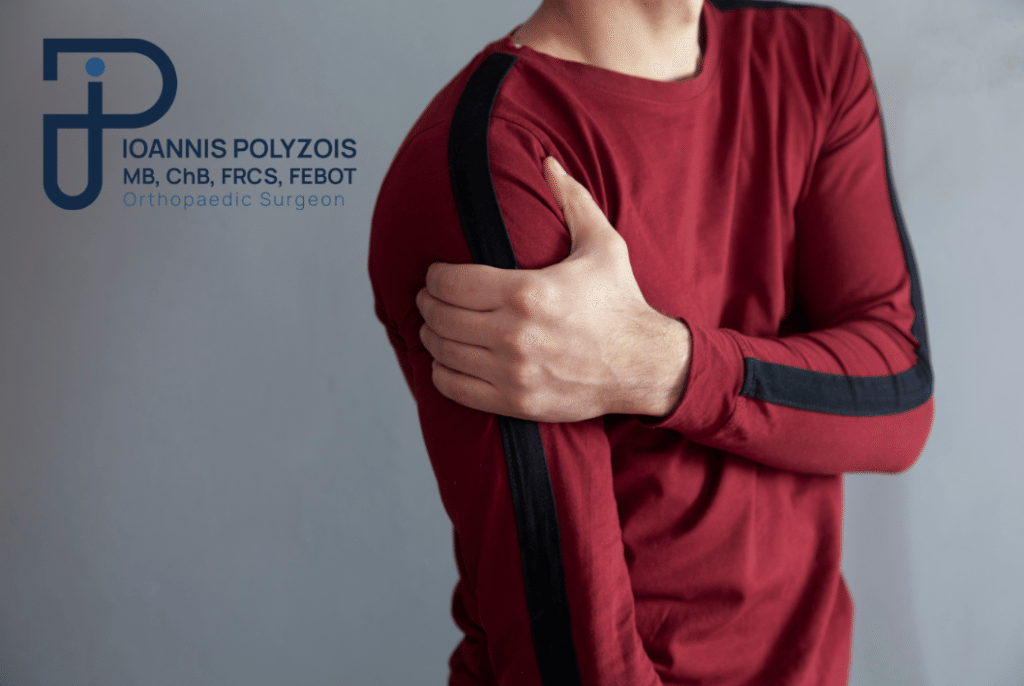
The Natural Course of the Disease: The 3 Stages of Frozen Shoulder
Frozen shoulder does not strike one day and go away the next. It is a condition with an extremely long and often distressing duration. If left untreated, the natural course of the disease passes through three very distinct stages, the total duration of which can exceed 2 or even 3 years.
First Stage: The Phase of Inflammation and Pain (Freezing Stage)
Duration: Usually 2 to 9 months.
Symptoms: This is the most painful stage. The inflammation of the capsule is at its peak. The patient experiences a deep, acute, and piercing pain that worsens day by day. The most characteristic symptom is the unbearable nocturnal pain, which destroys sleep and causes chronic exhaustion. Every sudden movement (e.g. reaching for the seat belt in the car) causes an “electric shock”. Stiffness begins to make its appearance.
Second Stage: The Phase of “Freezing” and Stiffness (Frozen Stage)
Duration: 4 to 12 months.
Symptoms: The paradox here is that the acute inflammation begins to subside and, consequently, the pain calms down (mainly at rest). However, the adhesions have now completely hardened. The shoulder is literally “frozen” and locked. The patient faces extreme difficulty with daily movements: they cannot comb their hair, put on a bra, wash their back, or put on a jacket. External and internal rotation of the arm have practically been lost.
Third Stage: The Phase of Resolution and Recovery (Thawing Stage)
Duration: 5 to 24 months.
Symptoms: This is the stage of natural “thawing”. The contraction begins to recede very slowly. The pain is minimal, and the mobility of the shoulder improves gradually, returning (in most, but not all cases) to normal or near-normal levels.
The critical question: Why should a patient wait 2 or 3 years experiencing pain and disability, when modern medicine can provide an immediate solution?
The Diagnostic Approach: Excluding Mistakes
The diagnosis of frozen shoulder requires clinical insight. Many conditions (such as a tendon tear or advanced arthritis) mimic the pain and stiffness. As a sceptical doctor, I must prove what it is not, to conclude what it is.
The Clinical Examination (The Key): The absolutely characteristic finding of true frozen shoulder is the simultaneous loss of both active and passive movement. If the patient cannot lift the arm by themselves (active), but I can lift it effortlessly for them (passive), then it is not frozen shoulder, but a tendon tear. Furthermore, the hallmark of frozen shoulder is the extreme loss of passive external rotation (the patient cannot open the arm outwards with the elbow stuck to the side).
- Simple X-ray: It is mandatory. Not to see the frozen shoulder (the capsule is not visible on X-rays), but to rule out severe osteoarthritis, osteophytes, or old fractures that cause stiffness. In a frozen shoulder, the X-ray is usually completely normal.
- Ultrasound and Magnetic Resonance Imaging (MRI): They are used to confirm the integrity of the tendons of the rotator cuff. On the MRI, an experienced doctor can distinguish the characteristic thickening of the inferior joint capsule and the swelling in the area of the rotator interval, confirming the diagnosis indisputably.
Conservative Treatment: The First Step Towards Release
Once we have established the accurate diagnosis, the planning of the treatment depends on the stage in which the patient is. In the early stages, the approach is always conservative.
- Anti-inflammatory Therapy: In the first, painful stage, strong non-steroidal anti-inflammatory drugs (NSAIDs) are administered to break the pain cycle, so that the patient can sleep.
Intra-articular Corticosteroid Injections: This is perhaps the most effective non-surgical intervention in the inflammation phase. The injection of cortisone directly inside the joint (usually under ultrasound guidance for absolute accuracy) dramatically reduces the swelling of the capsule and the nocturnal pain. In some cases, the injection of autologous biological factors (PRP) is also applied.
- Distension with Saline (Hydrodistension): A technique where we inject a large quantity of saline and local anaesthetic into the joint, with the aim of “inflating” the contracted capsule and partially breaking the adhesions from within.
- Specialised Physiotherapy: It is absolutely essential, but it must be done with great care. A frequent mistake is the application of excessive force (aggressive stretches) by the physiotherapist in the first (inflammatory) stage. Aggressive physiotherapy when the shoulder is “boiling” with inflammation causes micro-injuries that worsen the freezing. The stretches must be mild, controlled, and within the limits of pain tolerance.
Surgical Treatment: The Arthroscopic Revolution
In a significant percentage of patients (especially in diabetics), conservative treatment fails to break the hard adhesions. When the pain remains unbearable, the stiffness persists for more than 4–6 months without improvement, and daily life has become martyrdom, remaining on conservative means is simply a waste of time.
There, modern surgery intervenes offering the optimal, immediate, and impressive solution: Arthroscopic Capsular Release.
How is the operation performed?
As a proponent of minimally invasive techniques, I apply Shoulder Arthroscopy, which completely changes the experience of the patient. There are no large incisions, no blood.
- 2 or 3 microscopic openings are made in the skin, only 4 millimetres in diameter.
- A high-resolution camera (arthroscope) is inserted. The image of the frozen shoulder internally is characteristic: a deep red, thick capsule, full of fibrous bands that are strangling the joint.
Using specialised state-of-the-art instruments, such as special radiofrequency devices (RF wands), I proceed to a 360-degree controlled division (cutting) of all the pathological adhesions and of the contracted capsule. I fully release the rotator interval and the ligaments.
The Result is Immediate: Even while you are on the surgical table under anaesthesia, I check your shoulder. From complete “locking”, the shoulder recovers 100% of its free, normal movement in every direction, within 30–40 minutes!
The Advantages of the Arthroscopic Solution
- Absence of Postoperative Pain: In cooperation with the specialised anaesthesiologist of our team, advanced regional anaesthesia (interscalene block) is applied. This guarantees that you will wake without the slightest pain, and your arm will remain “numb” and pain-free for the next 12–24 hours.
- Immediate Mobilisation: There is no sling! On the contrary, it is required that you start moving your arm immediately.
- Day-case Hospitalisation: You are discharged and return home a few hours after the operation is completed.
- Safety: It is an extremely safe, bloodless operation with almost zero rates of complications in the hands of a specialised doctor.