Αρθροσκόπηση Ώμου

Shoulder Arthroscopy

ΠΕΡΙΕΧΟΜΕΝΑ

Shoulder pain is not merely a nuisance; it is a condition that can paralyse daily life, deprive you of sleep, and cancel out any professional or sporting activity. The shoulder joint is the most mobile in the human body, but this very freedom of movement makes it extremely vulnerable to injuries, wear, and degenerative changes. In modern Orthopaedic Surgery, the era of large, open, and painful surgical incisions is irrevocably over. Shoulder Arthroscopy is today the definitive, minimally invasive surgery method that we specialised Orthopaedic Surgeons use to inspect, to diagnose with absolute accuracy, and — most importantly — to definitively treat any pathology within the joint. As a doctor who relies on research, documentation, and detail, I do not approach any condition superficially. The purpose of this comprehensive guide is to provide you with every possible piece of information about shoulder arthroscopy, to resolve every question you may have, and to explain why this particular method is the top choice for your return to a normal life.


Ο μόνος Ορθοπαιδικός στην Ελλάδα στην χειρουργική ώμου και άνω άκρου με προϋπηρεσία μόνιμης διευθυντικής θέσης (substantive Consultant ) για 10 συναπτά έτη στην Μεγάλη Βρετανία

What exactly is Shoulder Arthroscopy?

Etymologically, the word comes from the ancient Greek words “arthron” (meaning joint) and “skopeo” (meaning to observe, to examine in depth). Literally, it means “to examine the joint from within”. In practice, shoulder arthroscopy is a technological marvel of medicine. During the operation, I insert a microscopic but powerful camera (the arthroscope), which features optical fibres and a lighting system, into the joint through an opening (portal) no more than 3–4 millimetres in diameter.

This camera, which supports High Definition (HD) or even 4K resolutions, projects the interior of the shoulder onto large surgical monitors. Due to the magnification and brightness, I can see the structures of the shoulder with crystal-clear detail — often with greater accuracy than that offered by the most modern Magnetic Resonance Imaging (MRI). Using these highly detailed images as a guide, I introduce through other microscopic openings specialised, fine surgical instruments (2 to 4 millimetres in diameter). With these instruments, I anatomically repair the damage, suture torn tendons, clean inflammation, and stabilise the joint. The result? We correct complex problems without injuring the normal tissues, ensuring immediate mobilisation.

Brief Anatomy of the Shoulder: What do we examine arthroscopically?

To understand the value of the operation, it is necessary to know what exists inside your shoulder. The shoulder joint (glenohumeral joint) resembles a golf ball (the head of the humerus) resting on a small tee (the glenoid of the scapula). Because this bony contact is shallow, stability depends entirely on the soft tissues:

  • The Rotator Cuff: A group of four tendons (Supraspinatus, Infraspinatus, Subscapularis, Teres Minor) that embrace the head and allow it to lift and rotate.
  • The Labrum: A ring of durable cartilage that deepens the socket of the glenoid and keeps the “ball” in place.
  • The Joint Capsule: The sac that surrounds the joint and secretes the lubricating fluid.
  • The Subacromial Space and the Bursa: The “cushion” that protects the tendons from friction with the acromion bone.
  • The Tendon of the Long Head of the Biceps: It passes through the joint itself and contributes to stability.

Through arthroscopy, we meticulously examine each one of these structures.

The Evolution and Advantages of the Arthroscopic Method

Shoulder arthroscopy has been performed since the 1970s, but in recent years, it has made tremendous progress. Modern radiofrequency tools, absorbable anchors, and ultra-high-strength sutures have changed the rules of the game.

The benefits for you, the patient, are non-negotiable:

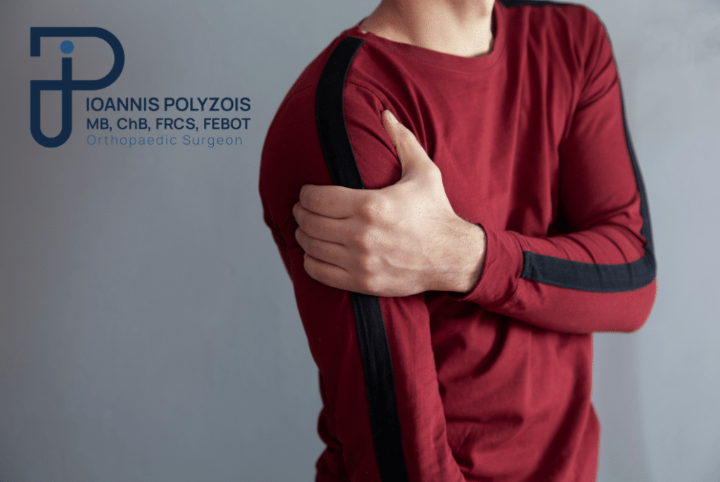
  • Bloodless and Painless Technique: There are no open incisions. Healthy muscles are not cut, injured, or violated. As a result, there is no bleeding, and the postoperative pain is minimal compared to the past.
  • Rapid Tissue Healing: The small openings (portals) lead to minimal tissue damage, allowing the body to focus its healing capacity on the lesion itself (e.g. on the sutured tendon) rather than on the skin wound.
  • Same-day Discharge (Day Clinic): Arthroscopy does not require lengthy hospitalisation. Patients, after a short monitoring period in the recovery room, return home a few hours after the operation is completed.
  • Safety and Accuracy: The camera provides visibility into the “blind” points of the joint, preventing medical errors and ensuring 100% anatomical correction.
  • Excellent Cosmetic Result: After a few months, the microscopic scars on the skin are often impossible to detect with the naked eye.

Which Shoulder Conditions are Treated Arthroscopically? (Detailed Presentation)

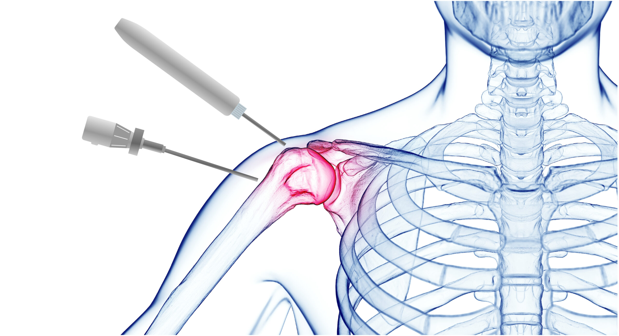
The technique of arthroscopy in the hands of a specialised doctor is not standardised; it is fully personalised. It addresses 95% of traumatic and degenerative shoulder conditions. Let us see in detail the most common:

Rotator Cuff Tears:

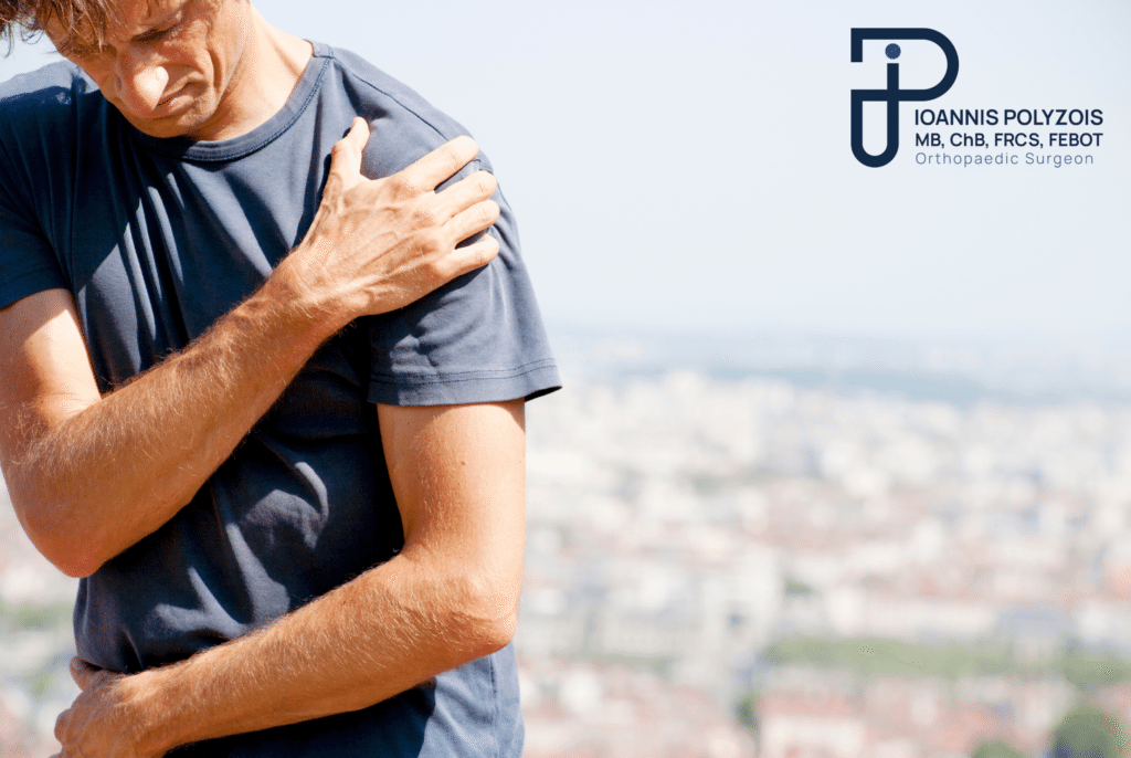
One of the most common causes of pain and weakness. The tendons of the rotator cuff, either due to injury (e.g. lifting a weight) or due to natural wear, tear and detach from the bone. The patient is unable to raise the arm and suffers unbearable pain at night. The Arthroscopic Solution: We insert special anchors (small implants of biocompatible material) into the humeral bone. These anchors carry strong sutures, with which we “catch” the torn tendon and fix it back to its anatomical position. This technique fully restores strength and mobility.

Impingement Syndrome & Shoulder Tendinitis:

The subacromial space is the “tunnel” through which the tendons pass. When the bone above (the acromion) develops “spurs” (osteophytes), the space narrows. Every time you lift your arm, the tendon rubs against (impinges on) the bone, causing inflammation, swelling, and intense pain. The Arthroscopic Solution (Acromioplasty): Using a special microscopic burr (shaver/burr), we “smooth out” the protruding bone, enlarge the tunnel, and remove the inflamed bursa. The tendon is immediately released.

Calcific Tendinitis:

For unknown reasons, often in middle-aged women, calcium crystals (like chalk or toothpaste) form within the mass of the tendon. This causes the most acute, dramatic pain in orthopaedics. The patient often goes to the emergency department unable even to move the arm. The Arthroscopic Solution: We pinpoint the exact location of the calcium deposit with the help of the camera, open the tendon, clean it, and aspirate all the material. The patient wakes up from the operation practically cured of the pain.

Frozen Shoulder (Adhesive Capsulitis):

The joint capsule thickens, becomes stiff, and fills with adhesions. The shoulder “locks” completely. It is a distressing condition that torments the patient for months or even years. The Arthroscopic Solution (Capsulotomy): Instead of waiting years for it to “thaw” on its own, we enter arthroscopically and, using a special radiofrequency instrument, we cut the adhesions and “open” the capsule 360 degrees. The shoulder is released at that very moment, and as early as the next day the patient has a full range of motion.

Shoulder Instability & Dislocations (Bankart Lesion / Hill-Sachs):

Common in young people and athletes. When the shoulder dislocates, it tears the labrum and the ligaments (Bankart Lesion). If not repaired, the shoulder will continue to dislocate (recurrent dislocation), gradually destroying the cartilage. The Arthroscopic Solution: We perform arthroscopic stabilisation. We place anchors in the glenoid and suture the torn ligaments back to their position, recreating the “protective wall” that keeps the shoulder stable.

SLAP Lesions (Superior Labrum Anterior and Posterior):

A tear at the upper part of the labrum, where the tendon of the biceps attaches. Very common in throwing athletes (tennis, volleyball, basketball). Treated with arthroscopic fixation of the lesion or biceps tenodesis.

The Sceptical, Scientific Approach: Preoperative Assessment

As a scientist who takes safety and effectiveness very seriously, I do not operate as a “surgery machine”. Every patient passes through the “filter” of absolute documentation. I never rely exclusively on a written report by a radiologist. I personally read and study the digital MRI files, double-checking the findings against your symptoms.

At our practice, the process begins with:

  • Taking an Exhaustive Medical History: I am interested in knowing exactly when you experience pain, which movements cause you difficulty, what medication you have taken, what are your work and sporting demands.
  • Detailed Clinical Examination: I perform special, targeted clinical tests of kinesiology and strength (e.g. Jobe’s test, Neer’s test, Apprehension test) to confirm the condition with my own hands.

If we conclude that arthroscopy is the indicated solution, a strict preoperative assessment follows for the safe execution of the operation. This includes:

  • Full blood and coagulation tests.
  • Electrocardiogram (ECG) and full cardiac assessment.
  • Chest X-ray.

In case there are underlying conditions (e.g. diabetes, hypertension, arrhythmias, or a history of infections), the experienced and specialised anaesthesiologist of our team proceeds to further evaluation. Your safety is non-negotiable.

Attention: It is of vital importance to inform us about every medication or dietary supplement you are taking. Especially for patients under anticoagulant therapy (such as Salospir, Plavix, Eliquis, Xarelto), precise instructions will be given for their temporary discontinuation or substitution before the operation.

The Procedure Step-by-Step: What happens in the Operating Theatre?

Once you arrive at the medical centre, having followed the instructions to be absolutely fasting, our team will welcome you to prepare you.

The matter of Anaesthesia:

Our anaesthesiologist will explain the options to you. In most shoulder arthroscopies, we prefer a combination of Regional (Local) and General or Light Sedation.

Initially, a “block” (Interscalene block) is performed. With the help of ultrasound, the anaesthesiologist injects local anaesthetic into the nerves leading to the shoulder. This means that your shoulder “falls asleep” and is completely numb. The huge advantage of this block is that it offers complete analgesia (zero pain) for 12 to 24 hours after the end of the operation! Then, light sedation or general anaesthesia is administered, so that you neither hear nor feel anything at all, ensuring your comfort.

The Surgical Technique:

  • Positioning: Once you are asleep, you are placed in a special chair (the “Beach Chair” position) or on your side, so that we have complete access to the joint from every angle.
  • Joint Distension: We inject sterile saline solution with a special controlled-pressure pump to “inflate” the joint and remove any blood, ensuring perfect visibility.
  • Diagnostic Tour: The first opening is made at the back of the shoulder. The camera is inserted. I meticulously check the cartilage, the tendons, the biceps, and the ligaments. I compare the image with the MRI and the clinical examination. Here, the action plan is confirmed or modified.
  • Therapeutic Intervention: I open 1–2 additional microscopic openings (at the front and side). Using specialised instruments (shavers for cleaning, radiofrequency for haemostasis, suture-passing instruments), I repair the damage. In tendon repairs, the latest-generation anchors are used, often bioabsorbable or knotless, which distribute the tension optimally.
  • Closure: The fluid is removed. The small openings close with a small adhesive strip (Steri-strip) or a single suture. There are no gauzes and no blood, only small waterproof plasters.

The surgical time, thanks to specialisation and technology, usually lasts from 20 to 45 minutes in most cases.

Meet the Doctor: Dr. Ioannis Polyzois

Medicine is not just a technique; it is responsibility, continuous research, and critical thinking. As an Orthopaedic Surgeon with absolute specialisation in conditions of the shoulder and upper limb, my primary goal is to offer you a safe, definitive, and scientifically documented solution. I do not experiment, and I leave nothing to chance.

Having served as a permanent Consultant in Orthopaedics in Great Britain (NHS) for more than 10 years, I was trained in one of the most demanding and strict healthcare systems in the world. My extensive further training at the largest centres of arthroscopic and reconstructive surgery worldwide enables me to apply the most modern international techniques in Greece.

To date, I have performed more than 9,000 arthroscopic and open surgical operations (of which more than 7,000 are purely arthroscopic). This vast surgical experience makes me competent for the successful execution of high-difficulty operations. Every patient is unique, which is why my approach is always entirely personalised. I am here to diagnose your problem with honesty and, together, to plan your return to normality.

Cost and Price: Shoulder Arthroscopy

A medical procedure is not a product with a “price tag”. The question about the cost of a shoulder arthroscopy is entirely reasonable, but the answer cannot be given over the phone or via the internet.

The reason is simple: The price is shaped according to the severity of the injury and the materials required. A simple arthroscopic removal of calcium deposits without the use of implants has a different cost from the repair of a massive rotator cuff tear that requires 3, 4, or 5 advanced bioabsorbable anchors. In addition, the cost is influenced by the choice of clinic and by the coverage you may have from EOPYY or your private insurance.

At our practice, we work with absolute transparency. The exact cost of your particular operation is determined and discussed honestly only after the clinical examination and the review of your tests during our scheduled appointment.

Movement is life!

There is no reason to tolerate restriction, stiffness, and daily pain. The solution is scientifically feasible, bloodless, and safe. Contact our practice today to schedule an appointment. We will thoroughly study your findings, I will resolve every question of yours with absolute honesty, and together we will organise the personalised orthopaedic care that you deserve.

Η κIνηση εIναι ζωH!

Επικοινωνήστε με τον γιατρό για εξειδικευμένη ορθοπαιδική φροντίδα, προσαρμοσμένη στις ανάγκες σας

Postoperative Course: Recovery and Physiotherapy

As I always emphasise to my patients: To achieve an excellent outcome, with 100% mobility and strength, 50% depends on the operation and the other 50% on the rehabilitation. I am categorical about this.

Recovery is very fast, but it requires discipline.

  • Day 0: After recovery from anaesthesia, you rest in your room. The pain is minimal thanks to the nerve block. I visit you, we discuss the findings, we look together at photographs from inside your shoulder, and a few hours later… you go home.
  • First Days: Depending on the condition, you may wear a light arm sling (e.g. if we have sutured tendons). If we simply cleaned out calcium deposits or adhesions, you would move your arm freely from the very first day!
  • Physiotherapy: The programme is 100% personalised. There is no “one-size-fits-all recipe”. I maintain a network of top, specialised physiotherapists throughout Greece. I communicate directly with them, and we design your rehabilitation programme. Initially, we focus on passive movement (so that the joint does not freeze), then on active movement, and finally on strengthening.

Are there Complications? The Truth About the Risks

In science, there are no dogmas and guarantees, but there is data. Shoulder arthroscopy, when performed by an appropriately trained and experienced Orthopaedic Shoulder Surgeon, has an almost zero complication rate.

The risks of infection are minimal (less than 0.5%) thanks to the small incisions and the abundant saline solution that continuously flushes the joint. Risks of damage to nerves or vessels are eliminated by deep anatomical knowledge and the clarity of the camera. In general, it is one of the safest, most bloodless, and most effective operations in all of medicine.

Συχνές ερωτήσεις

What is the recovery time after shoulder arthroscopy?

The answer depends on the nature of the condition. If the operation involved cleaning, release of adhesions, or removal of calcium deposits, functionality returns dramatically within 1–2 weeks. If we performed a tendon repair (supraspinatus tear) or stabilisation of a dislocation, approximately 4–6 weeks of postoperative protection in a sling are required, and in total, 3 to 6 months of physiotherapy for full sporting or heavy manual return.

Are there complications after shoulder arthroscopy?

As previously mentioned, complications (such as infections, neurological injuries, or thromboses) are statistically minimal to zero, especially when the operation is performed by a strictly specialised doctor in a modern, certified surgical environment. It is, by definition, a safe operation.

Is physiotherapy necessary after shoulder arthroscopy?

Necessary. Without proper physiotherapy, the joint may develop stiffness, and the muscles may atrophy. The role of the physiotherapist is to “re-educate” your arm, to ensure the correct healing of the tendons, and to restore muscular balance around the scapula.

Is it normal to have pain after shoulder arthroscopy?

A mild, controlled pain or a sense of “heaviness” during the first 48–72 hours is absolutely normal, as the body reacts to the surgical intervention. However, thanks to the regional anaesthesia block and the painkilling regimen we prescribe for you, this pain is fully manageable and in no way resembles your preoperative pain.

What is the recovery time after general (open) shoulder surgery?

The “open” operations (e.g., arthroplasties, fractures) require a longer time for the healing of the muscles that have been divided. By contrast, arthroscopy reduces the recovery time by half.

How long does the recovery last after shoulder arthroscopy?

Recovery is a dynamic process. In 3 days, you will feel comfortable at home. In 2 weeks, the sling often comes off. In 4–6 weeks, you can drive. The full “maturation” of the tissues is completed at 6 months.

What exercises are performed after shoulder arthroscopy?

We initially start with pendulum-type exercises (Codman) and passive assisted movement (e.g., with pulleys or a stick). Then, isometric exercises (contraction without joint movement) are introduced, and in the final stage, exercises with resistance bands, light weights, and plyometric proprioception exercises are introduced.

When can I take a bath after the operation?

Thanks to the waterproof adhesive strips, you can shower as early as the second day, taking care not to rub the area of the incisions. Full submersion in water (bath, sea) is usually allowed after 10–14 days.

Will I have to give up sport?

The exact opposite. The purpose of arthroscopy, especially in athletes (professional and amateur), is the return to the field at the pre-injury level. The synchronisation and patience in physiotherapy are the “key” to this return.